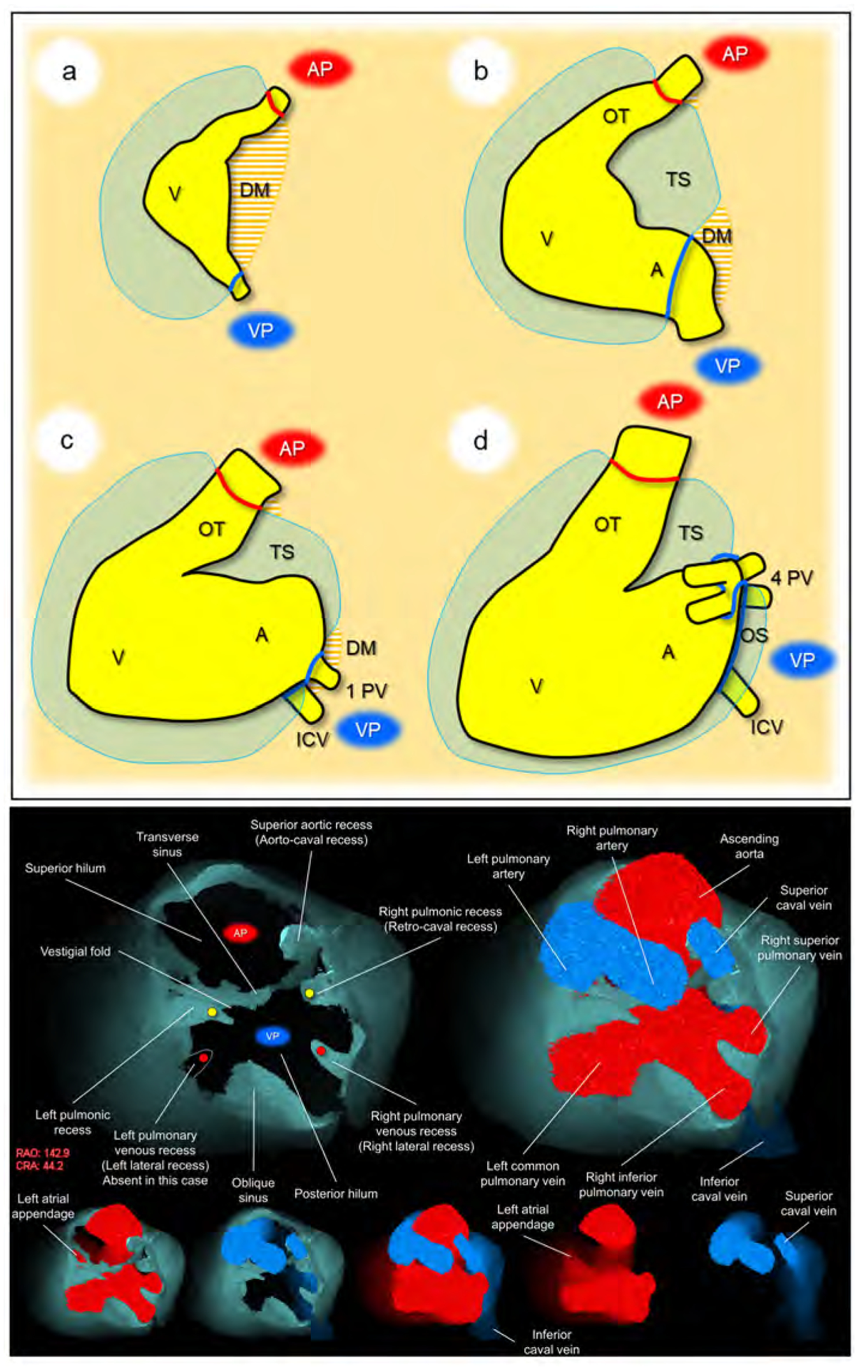
Figure 2. Development and living anatomy of the pericardial space.

Developmental concepts of the pericardial space (upper panel). a. The heart tube is anchored to the body wall by both arterial and venous poles and dorsal mesocardium. b. The dissolution of the dorsal mesocardium creates the transverse sinus. c. Single pulmonary vein developed from the mesocardium at the venous pole. d. Superior expansion of the four pulmonary veins creates the deep oblique sinus. Lower panel. Basal superior views of the superior hilum and posterior hilum without heart (left) and with heart (right). Lower five images show the composite and component images viewed from the same direction to show the detailed anatomical information. Pericardial space is reconstructed as the solid structure. Yellow circles indicate bilateral pulmonic recess. Red circles indicate bilateral pulmonary venous recess, although it should be noted that this patient does not show a prominent left pulmonary venous recess due to the left common pulmonary vein. Note the symmetry in these recesses. Vestigial fold, between the transverse sinus and left pulmonic recess corresponds to the ligament of Marshall, subsequent to regression of the left superior caval vein. Refer to Supplementary movie 1. A, atrium; AP, arterial pole; CRA, cranial; DM, dorsal mesocardium; ICV, inferior caval vein; OS, oblique sinus; OT, outflow tract; PV, pulmonary vein; RAO, right anterior oblique; TS, transverse sinus; V, ventricle; VP, venous pole.
