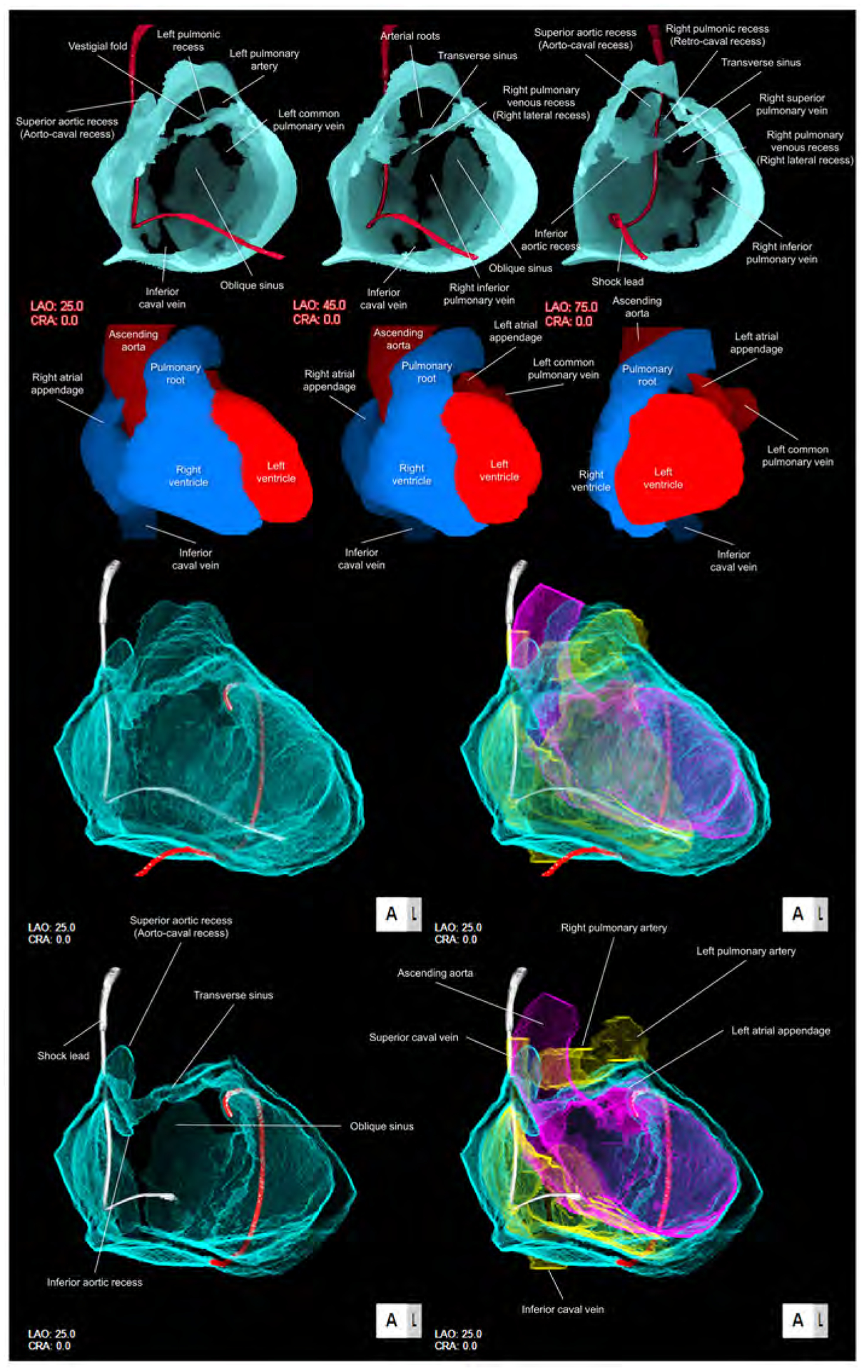
Figure 3. Solid and double-layer reconstructions of the pericardial space.

Upper panels are solid reconstructions of the pericardial space with corresponding cardiac chamber endocast images. Note that the course of the shock lead represents the location of the superior caval vein between the transverse sinus and right pulmonic recess. Also, the transverse sinus is continuous with superior and inferior aortic recesses. The narrowest diameter of the transverse sinus in this case is 1.4 × 1.8 mm. Refer to Supplementary movie 2. Oblique sinus is a tongue-like shaped blind end with its orifice demarcated by the inferior caval vein and the left inferior pulmonary vein. Lower panels are double-layer reconstruction of the pericardial space without (left panels) and with (right panels) heart shell. The right and left heart are colored in yellow and purple, respectively. Lower panels are sectioned to show the transverse sinus and oblique sinus in relation with cardiac chambers. Note the posterior course of the transverse sinus relative to the arterial roots. Oblique orifice of the oblique sinus is well observed in left lower panel. A drainage tube located adjacent to the left atrial appendage via the inferior course is reconstructed in red. Refer to Supplementary movies 3, 4 and 8. CRA, cranial; LAO, left anterior oblique.
