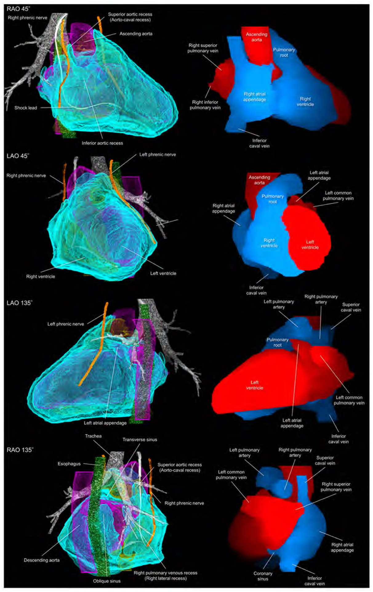
Figure 4. Living anatomy of the pericardial space and surrounding structures.

Pericardial anatomy with internal and external adjacent structures visualized in every 90° with corresponding cardiac chamber endocast images. In the left images, the right and left heart are colored in yellow and purple, respectively. The vertebral column and pleura are additionally reconstructed in the Supplementary movie 5. Refer to Figure 1. LAO, left anterior oblique; RAO, right anterior oblique.
