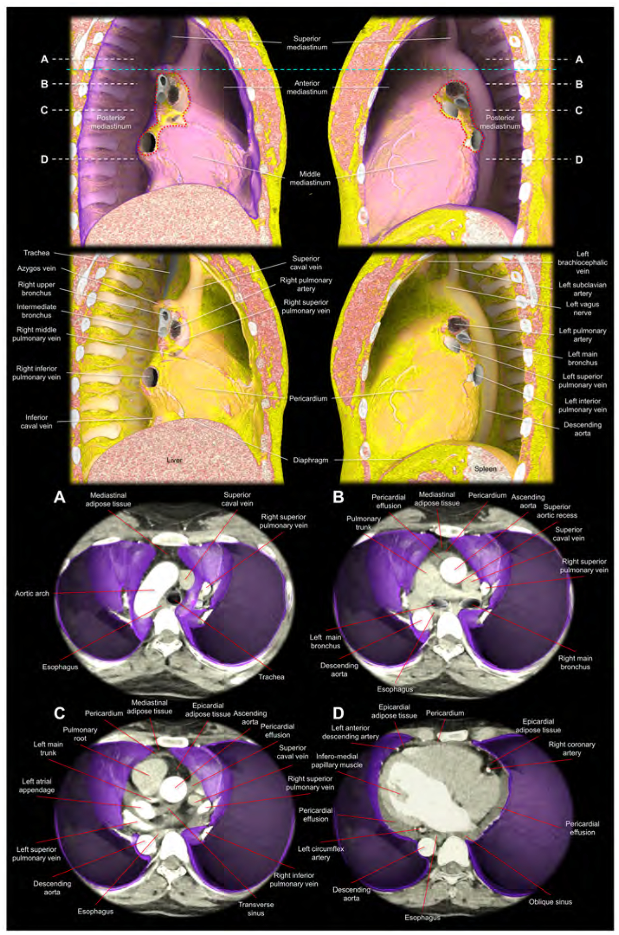
Figure 5. Virtual dissection images showing the relationship among the pericardium, parietal pleura, and mediastinal structures.

Images are reconstructed from the cardiac computed tomographic datasets obtained from a 35-year-old woman with pericardial effusion, which makes it easier to discern the pericardium corresponding to the outer margin of the pericardial effusion. The upper panels show the right and left lateral images of the thorax after virtual resection of the bilateral lungs. The purple region indicates the parietal pleura. The red dotted lines correspond to the estimated location of the pleural reflections surrounding bilateral pulmonary hila. Note the difference in the spatial relationship between the bronchus and pulmonary artery, when comparing the right and left pulmonary hila (65). The sky-blue dashed line corresponds to the horizontal plane sectioned at the height of the sternal angle used to define the superior mediastinum. The second upper panels show the additional virtual peeling off of the parietal pleura. The rich mediastinal adipose tissues located in the superior and anterior mediastinum are noted. Note the intercostal arteries, left vagus nerve, and the azygos vein overriding the right superior pulmonary vein, right pulmonary artery, and the right bronchus. Lower panels are virtual dissection images viewed from the superior direction after virtual resection of the bilateral lungs. Therefore, the bottom of the thorax corresponds to the diaphragmatic surface covered by the parietal pleura (purple). Panels A-D correspond to the planes A-D indicated in the upper panels (white dashed lines). Panel A shows the superior mediastinum, above the level of the anterior pericardial reflection. Thus, the adipose tissue anterior to the aortic arch is not the epicardial adipose tissue, but the mediastinal adipose tissue. Panel B shows the level just inferior to the superior margin of the anterior reflection of the pericardium. The adipose tissue anterior to the pericardium is the mediastinal adipose tissue. At the middle mediastinum, substantial part of the lateral pericardium is adjacent to the parietal pleura. Thus, the phrenic nerves, descending anterior to the both pulmonary hila, are sandwiched between the pericardium and medial part of the parietal pleura. Panel C shows the level of the transverse sinus. Rich epicardial adipose tissue, which is located interiorly to the pericardial space, can be observed. Panel D is the level of the infero-lateral papillary muscle of the left ventricle. Note the pericardial effusion filling postero-lateral pericardial space and the oblique sinus. From the pleural space, the pericardial space is accessible by penetrating the parietal pleura and the pericardium. Note the epicardial adipose tissue at the anterior interventricular groove and the atrioventricular grooves, which involve coronary arteries. Note the mediastinal adipose tissue of the anterior mediastinum behind the sternum becomes thinner in panel C compared to panel B, and it is almost undetectable in the panel D. The esophagus is distant from the left parietal pleura with the intervening of the descending aorta.
