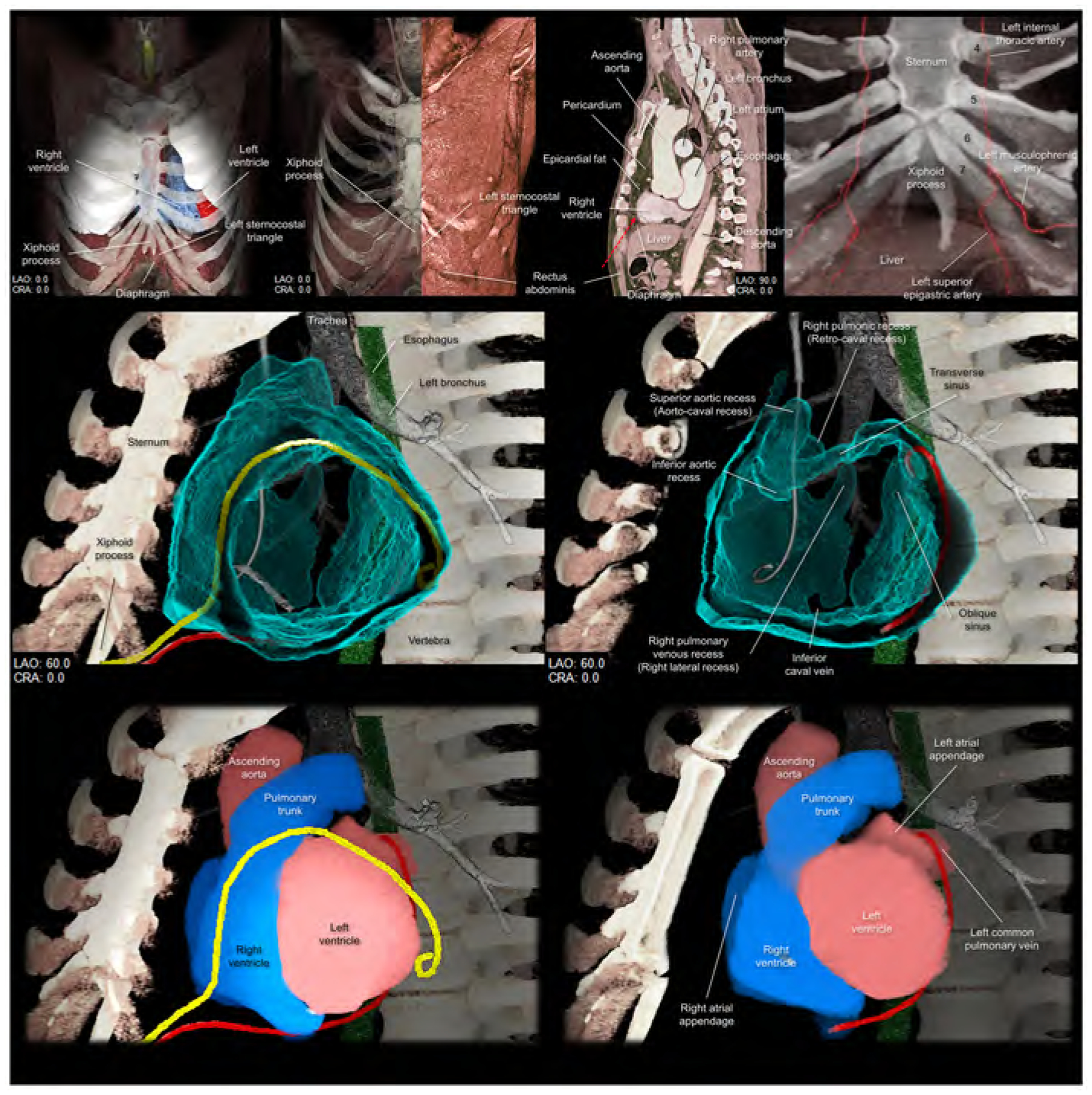
Figure 6. Structural anatomy around the left sternocostal triangle and subxiphoid approach.

Upper panels focus on the left sternocostal triangle. Left panel shows the frontal view demonstrating the relationship between the xiphoid process, diaphragm, and left sternocostal triangle. Left middle panel shows the rectus abdominis at the left sternocostal triangle. Right middle panel shows the sagittal plane, which is sectioned along the vertical line indicated in the middle panel, demonstrating the relationship between liver, rectus abdominis, and pericardium, and epicardial fat. Red dotted arrow indicates the direction of the epicardial access via the left sternocostal triangle, also called Larrey’s approach. Right panel demonstrates the anatomy of the left superior epigastric artery. Numbers in this panel indicate the costa. Middle panels are virtual dissection images to show the anterior and inferior course of the drainage tube (yellow and red, respectively) via the left sternocostal triangle. Red drainage tube inserted close to the left atrial appendage is reconstructed from the real tube, whereas the yellow drainage tube inserted close to the inferolateral region of the left ventricle is the virtual one. Lower panels are the images viewed from the same directions showing the spatial relationships between the drainage tubes and cardiac chambers. Refer to Supplementary movie 8. CRA, cranial; LAO, left anterior oblique.
