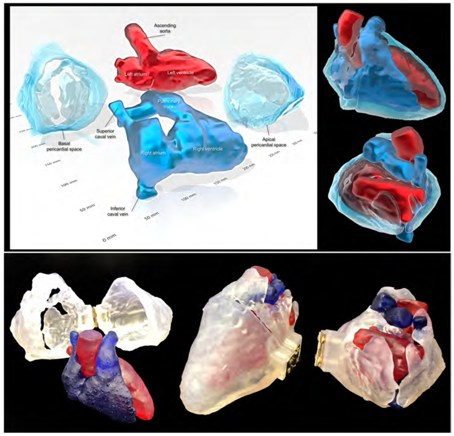
Figure 8. Three-dimensional prints of the pericardial ‘space’.

Images reconstructed from the STL file to create three-dimensional printing models (upper panels) using a commercially available software (3D Builder, Microsoft Co. Redmond, WA, USA). Lower panels show real three-dimensional printed models (using the 3MF file that is uploaded with this paper). Refer to Supplementary movie 9, Supplementary 3MF file, and Supplementary three-dimensional PDF file.
