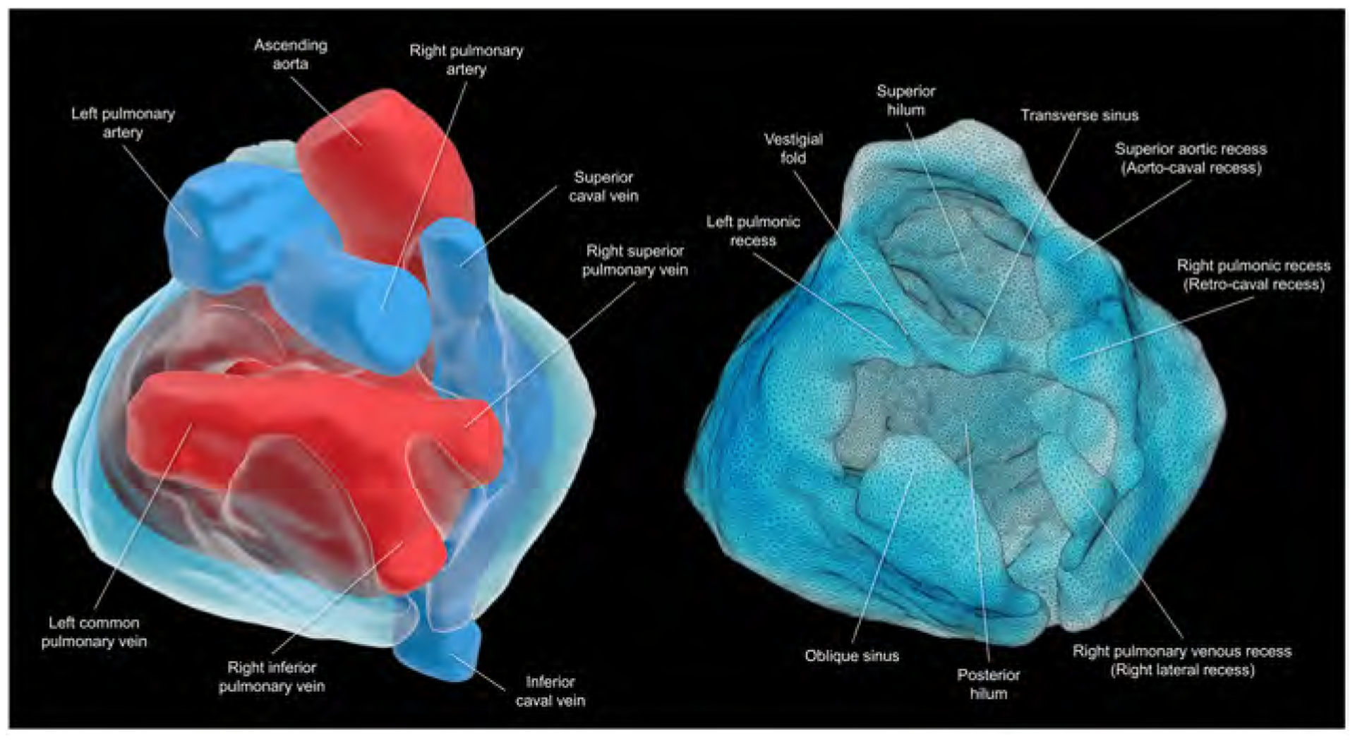
Central illustration. Living anatomy of the pericardial space.

Views of the superior hilum and posterior hilum with (left) and without (right) heart. Pericardial space is reconstructed as the solid structure. The superior hilum involves the ascending aorta and pulmonary trunk. The posterior hilum involves pulmonary veins and caval veins, separated by the transverse sinus. Both hila, separated by the transverse sinus, are the only entry/exit for the extracardiac nerves and vessels. This patient does not show prominent left pulmonary venous recess due to the left common pulmonary vein.
