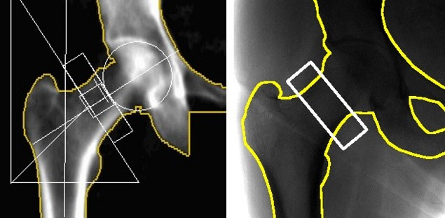
Figure 1.

(Left) Dual-energy X-ray absorptiometry image with the standard hip geometrical markings overlaid (white) and the bone-soft tissue boundary (yellow). (Right) Digital radiograph scan with as close as achievable match to hip placement. The ROI (white) shows the region used for femoral neck bone health analysis.
