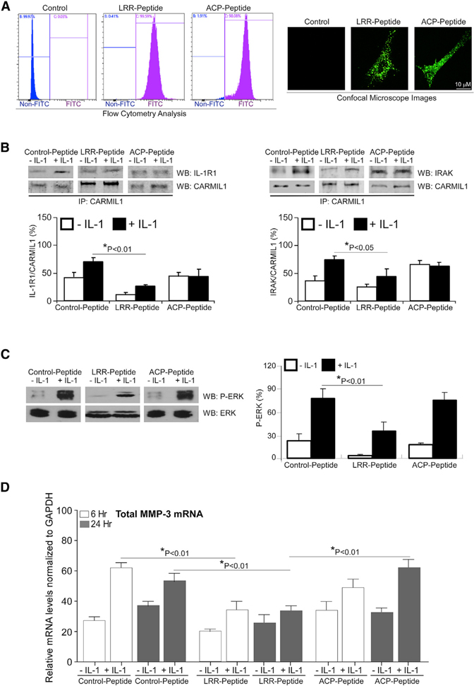
Figure 4. Effect of Cell-Permeable CARMIL1 Peptides on IL-1-Induced ERK Activation and MMP3 Expression.
(A) HGFs were incubated with FITC-labeled, cell-penetrating TAT LRR peptide (1 μM/mL) and CP peptide (1 μM/mL) overnight and analyzed by flow cytometry and viewed by fluorescence microscopy.
(B) HGFs transduced with FITC-labeled, cell-penetrating TAT control peptide, LRR peptide, and CP peptide were treated without or with IL-1 (20 ng/mL) for 45 min. CARMIL1 IPs were prepared from whole-cell lysates of the cells above and immunoblotted for IL-1R1 and CARMIL1 (left), and for IRAK and CARMIL1 (right). The histograms show the percent mean ratios of densities ± SE of IL-1R1 to CARMIL1 or of IRAK to CARMIL1, respectively. Data from three independent experiments were averaged, and the mean ratios ± SE (n = 3 replicates) are indicated.
(C) Whole-cell lysates from transduced cells in (B) treated without or with IL-1 (20 ng/mL) for 45 min were immunoblotted for p(T202/Y204)-ERK and total ERK. Ratios of p(T202/Y204)-ERK to ERK were quantified by densitometry and indicated as the percent mean ratios of densities ± SE of 3 replicates from 3 separate experiments.
(D) MMP3 mRNA levels were quantified in the aforementioned transduced cells that were treated without or with IL-1 (20 ng/mL) for 6 h and 24 h by qRT-PCR analysis, respectively. Values are means ± SE from n = 3 independent experiments.

