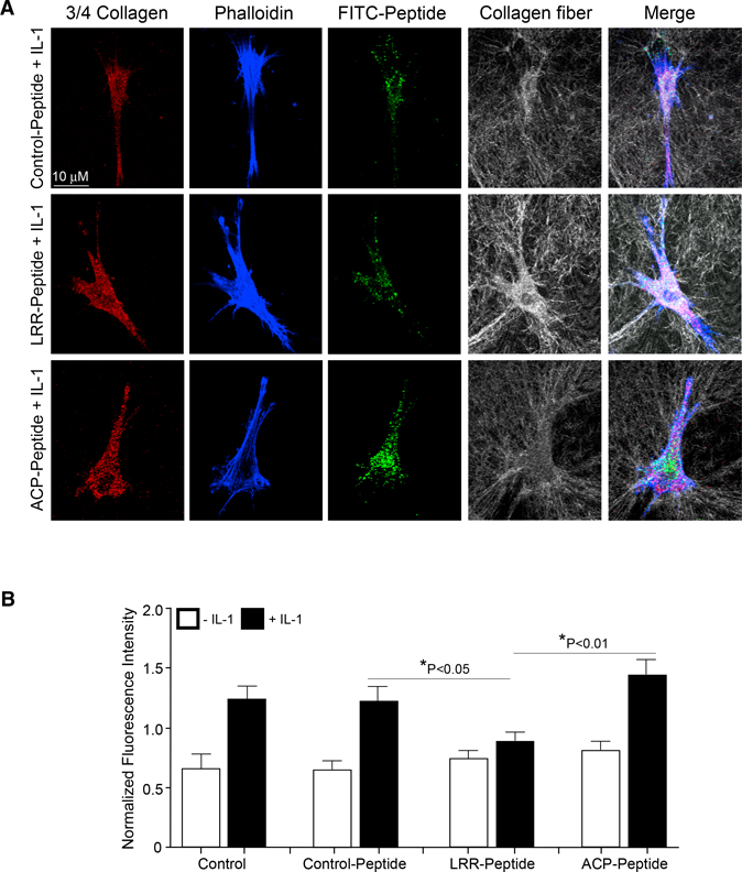
Figure 5. LRR Domain of CARMIL1 Is Associated with Inhibition of Collagen Degradation.
(A) Staining for 3/4 collagen fragments (red), actin filaments (phalloidin; blue), FITC-labeled peptide (green), and intact collagen fibrils (gray) by confocal reflectance (CR) in HGFs and HGF cells transduced with control peptide, LRR peptide, and CP-peptide plated on 3D, type I collagen gels and treated without or with IL-1 (20 ng/mL).
(B) Quantification of collagen degradation stained with a 3/4 collagen neoepitope antibody in the different treatments and conditions mentioned above. 3/4 collagen intensity levels were normalized to the overall average of all intensity values derived with ImageJ using 3/4 collagen signals. Values are means ± SE from n = 3 independent experiments.

