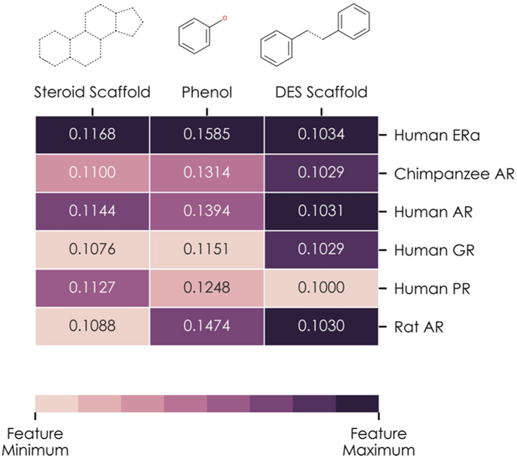
Figure 5.
Analysis of three chemical fragments known to bind to nuclear ERα and ERβ: the steroid scaffold, phenol group, and DES scaffold. Each column of the heatmap represents one chemical fragment and shows the weights of edges connecting the neuron associated with that fragment and each of the neurons in the network’s receptor binding layer: human ERα; chimpanzee, human, and rat AR; the human glucocorticoid receptor; and human PR. Each column is normalized such that the highest weighted edge leaving the fragment’s neuron is shown as the darkest color, and the lowest weighted edge is the lightest color.

