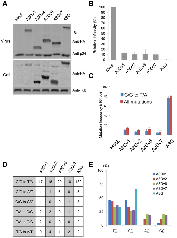
Figure 3. Antiviral activity of A3D and its variants on HIV-1.
(A) We transfected pNL4-3/Δenv/Δvif-Luc and pVSV-g with expression vectors for APOBEC3D variants and concentrated viral supernatant by ultracentrifugation. Viral concentrates and cell lysates were subjected to immunoblotting using indicated antibodies. Tub: Tubulin (B) Luciferase activity of HIV in the presence of APOBEC3D variants or APOBEC3G was measured with a luminometer and presented as relative values normalized to the value without APOBEC3 expression in the producer cells. Values represent the average of three independent experiments and error bars indicate S.D. (C) Mutation frequencies in HIV-1 gag PCR products derived from infected cells. 5 clones (2500 bps) were sequenced in each group. C/G to T/A mutation frequencies (blue bar) and mutation frequencies including all patterns (red bar) are shown. Error bars indicate S.E.M. (D) Mutation patterns of edited HIV-1 gag sequences. 5 clones (2500 bps) were sequenced for each group. (E) Dinucleotide patterns in edited HIV-1 gag. The rates of indicated dinucleotide sequence at C/G to T/A mutations.

