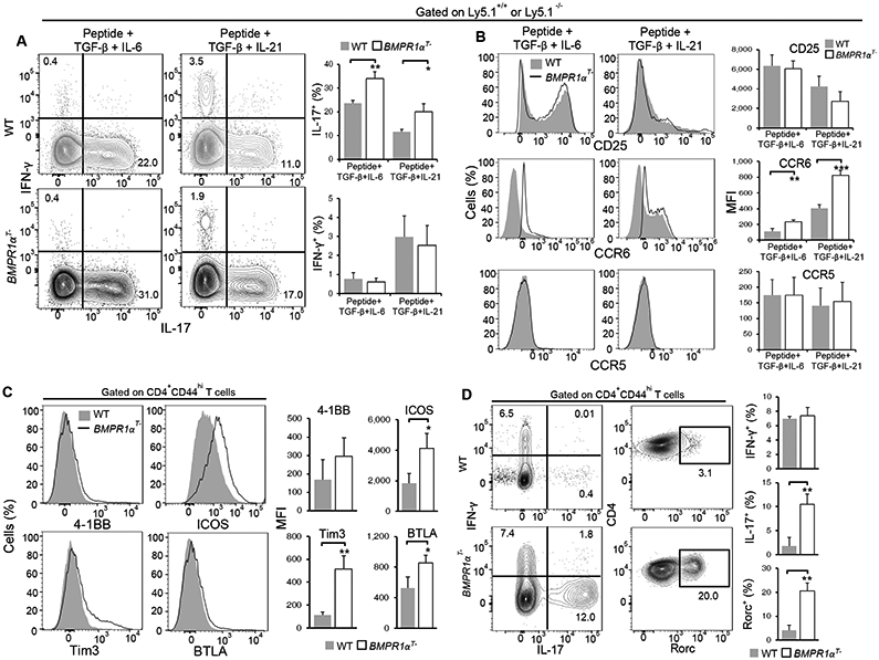
Fig. 4. Cell-intrinsic BMPR1α expression suppresses the generation of Th17 CD4+ T cells.

(A and B) Flow cytometry analysis of IFN-γ and IL-17 production (A) and surface marker expression (B) on co-cultured TCR transgenic WT and BMPR1αT− CD4+ T cells stimulated with antigenic peptide and the indicated cytokines for 6 days. Contour plots and histograms (left) are representative of three independent experiments. The frequency of cytokine producing cells and marker mean fluorescence intensity (MFI) (right) are means ± SD from all experiments. (C and D) Flow cytometry analysis of surface marker expression (C), IFN-γ and IL-17 production (D) and Rorc abundance (D) in lymph node T cells isolated 5 days after TCR transgenic WT and BMPR1αT− mice were immunized with antigenic peptide and CFA. Histograms and contour plots (left) are representative of three independent experiments. The frequency of cytokine producing cells and marker MFI (right) are means ± SD from all experiments. *P< 0.05, **P< 0.01, ***P< 0.001, as determined by Student’s t-test.
