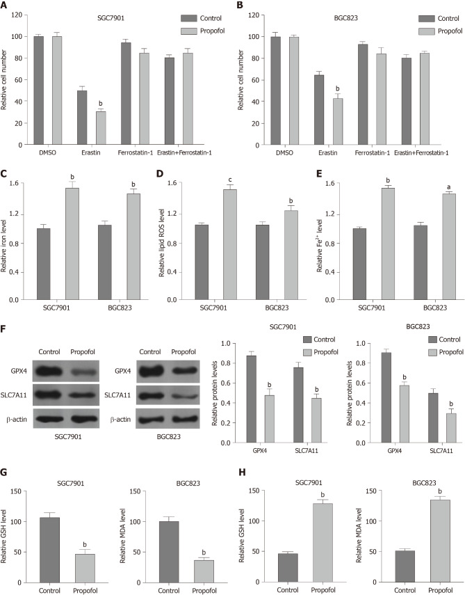
Figure 3.
Propofol enhances ferroptosis in gastric cancer cells. A and B: SGC7901 and BGC823 were cotreated with 5 mmol/L erastin or ferrostatin (1 mmol/L) and propofol (10 µmol/L). Cell growth was analyzed by MTT assays. C–F: SGC7901 and BGC823 cells were treated with propofol (10 µmol/L). C: Flow cytometry measured the levels of ROS. D and E: Iron Assay Kit analyzed the levels of iron and Fe2+. F: Western blotting analysis tested the expression of GPX4, SLC7A11 and β-actin. G and H: Levels of GSH and MDA were analyzed by the detection kit. n = 3, mean ± SD, aP < 0.05, bP < 0.01, cP < 0.001.

