Fig. 7.
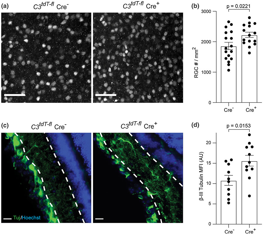
C3 depletion from astrocytes protects RGC and neurite projections in the retina. a Representative images of Brn3a staining in flat-mount whole retina from C3tdT–fl x GFAP-Cre mice at EAE PID42. Magnification 20x; Scale bar = 50 μm. b. Quantification of Brn3a + RGC number from different groups at PID42. c Representative images of Tuj staining in the retina of C3tdT–fl x GFAP-Cre mice at EAE PID42. Magnification 40x; Scale bar = 20 μm. d Quantification of Tuj staining in C3tdT–fl x GFAP-Cre mice with EAE, presented as MFI. Error bars represent SEM. AU arbitrary unit

