
Fig. 6. Cell studies to investigate PRA:PRB ratio and myometrial contractility.
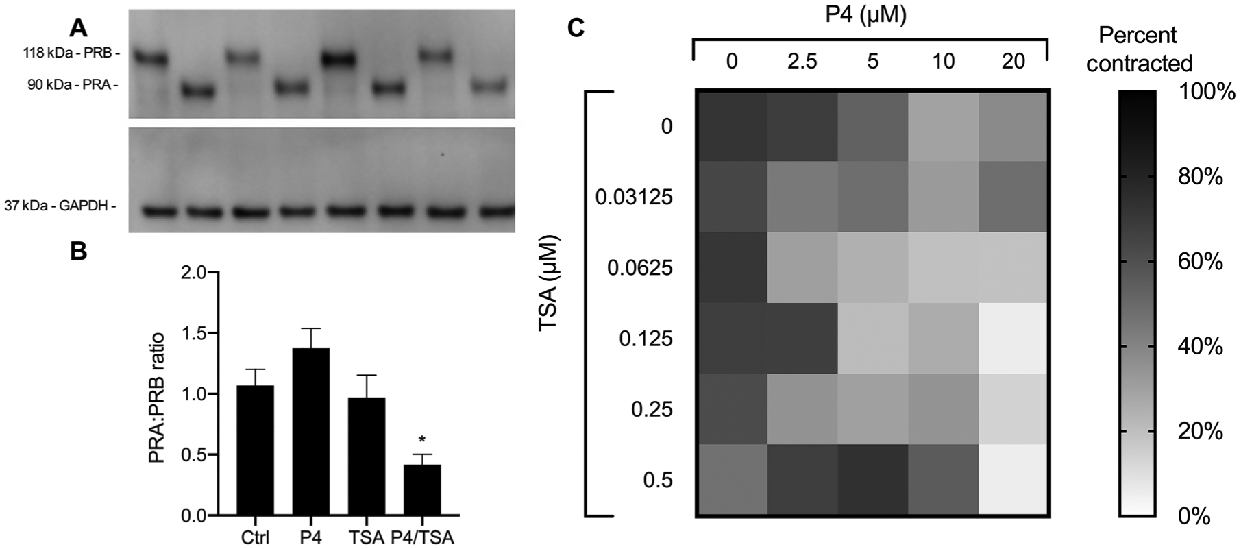
(A) Representative Western blot showing stability of induced PRA and PRB in hTERT-HMA/B cells cultured in the presence of P4, TSA, or P4/TSA for 24 hours. GAPDH, glyceraldehyde-3-phosphate dehydrogenase. (B) Quantification of PRA:PRB ratio. Data are shown as means ± SEM (n = 3). *P < 0.05 compared to untreated cells (Ctrl). (C) Contraction of PHM1–41 cells in a collagen gel after incubation with P4 and TSA at various concentrations. The percentage of contraction was calculated at 96 hours based on gel area relative to the negative control (empty well, 0% contraction). Data are presented as the means (n ≥ 6).