
Fig. 4. CSPG4.CAR-Ts do not cause tumor escape due to antigen loss.
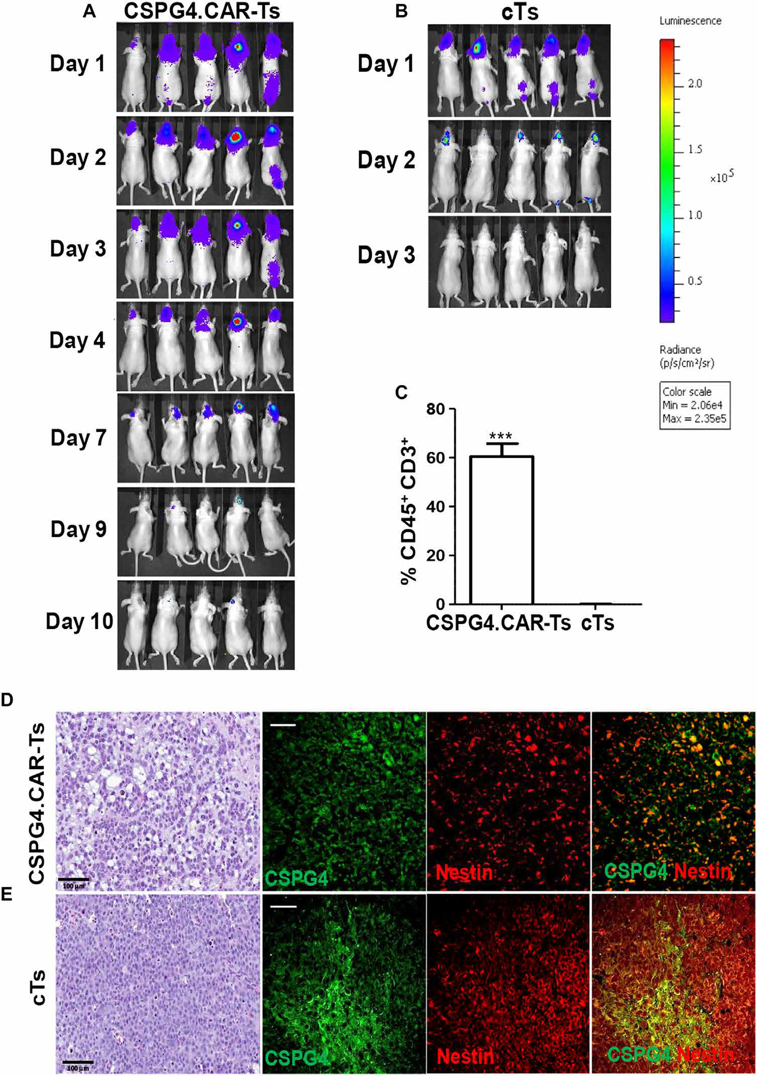
(A and B) Nude mice implanted intracranially with GBM-NS CSPG4H (96%, BT308-NS) were infused intratumorally with either CSPG4.CAR-Ts (A) or cTs (B) labeled with GFP/FFluc. Bioluminescence imaging shows the persistence of CSPG4.CAR-Ts (A) and cTs (B). (C) Bar graph showing human T cells detected intratumorally in xenograft gliomas removed from mice treated with CSPG4.CAR-Ts or cTs. Human CD45+CD3+ T cells were detected by flow cytometry. (D and E). Immunohistochemistry and immunofluorescence of GBM-NS CSPG4ML (49%, BT168-NS) glioma removed from mice previously treated with either CSPG4.CAR-Ts (D) or cTs (E). Hematoxylin and eosin staining of gliomas from mice treated with CSPG4.CAR-Ts shows a disruption in the architecture as compared to glioma from mice treated with cTs. Immunofluorescence of gliomas shows that CSPG4 and nestin expression is highly preserved in gliomas treated with either cTs or CSPG4.CAR-Ts (scale bars, 50 μm).