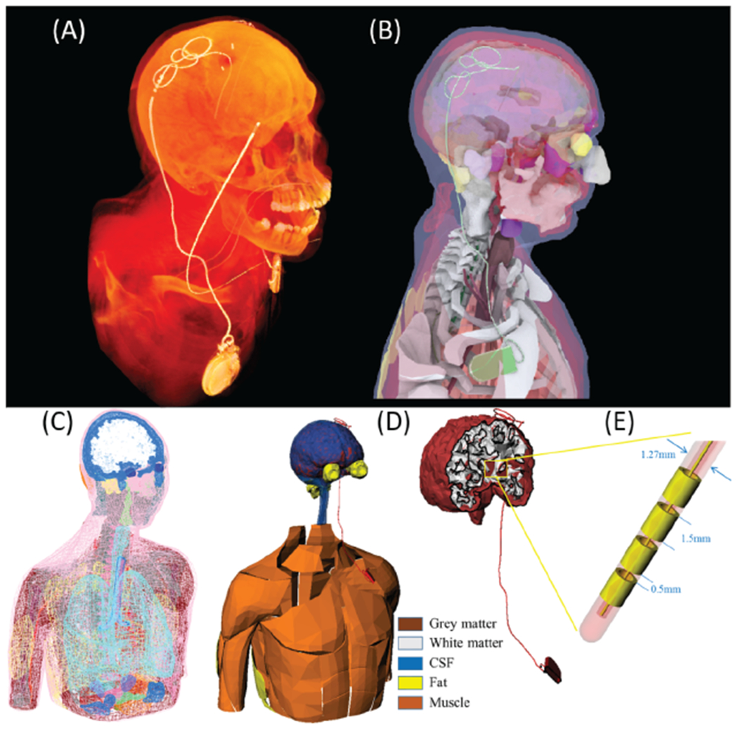
Figure 2.

(A) and (B) CT image of a patient with an implanted DBS implant. Device trajectory was manually segmented and a 3D model of the implant was constructed and registered to a heterogeneous human body model consisting of 184 individual body parts categorized into 30 tissue classes. (C) and (D) Conductivity of five major tissues surrounding DBS device was varied by ± 40% around their nominal values while other tissue conductivities were kept constant. (E) DBS lead model consisting of 4 electrode contacts, a hollow insulation, and a straight core.
