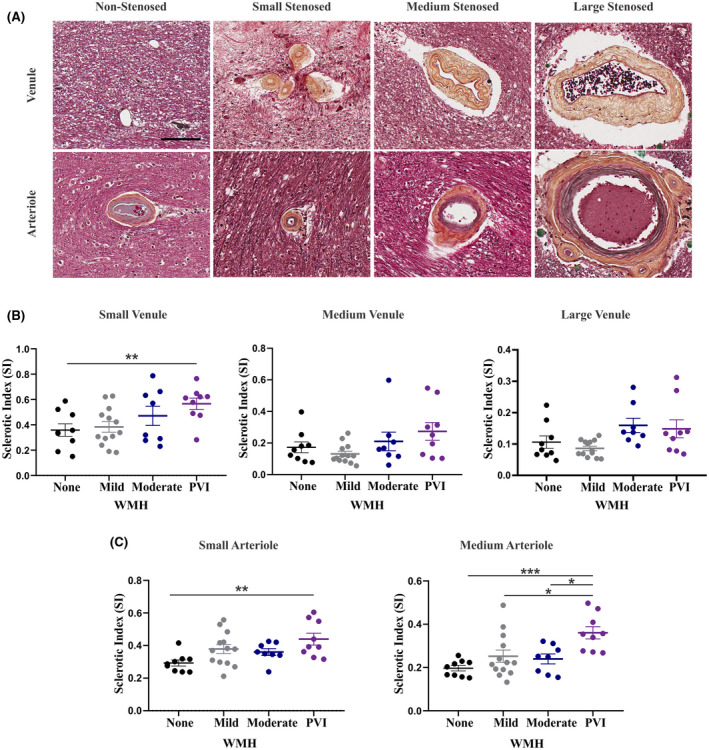
FIGURE 2.

Stenosis of both the small arterioles and venules is evident in the periventricular and subcortical white matter, and the severity is associated with imaging evidence of PVI. (A) Movat's pentachrome staining of arterioles and venules including non‐stenosed (with minimal collagen accumulation), small stenosed (<50 μm diameter), medium stenosed (50–200 μm diameter), and large stenosed (>200 μm diameter). All images captured at 20x magnification; scale bar indicates 100 μm. IHC for SMA was used to distinguish arterioles from venules. (B) Average SI of the small, medium, and large venules quantified in the periventricular white matter of each tissue block in specimens with none, mild, or moderate pvWMH or PVI. (C) Average SI of the small and medium arterioles quantified in the periventricular white matter of each tissue block in specimens with none, mild, or moderate pvWMH or PVI. SI of the small arterioles is increased in specimens with PVI in comparison to those with no WMH (p = 0.0063).SI of the medium arterioles is increased in specimens with PVI in comparison to those with no (p = 0.0006), mild (p = 0.0175), or moderate pvWMH (p = 0.0180). Comparison performed using one‐way ANOVA with Tukey's post‐hoc analysis, significance value of p = 0.05
