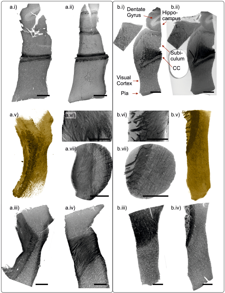
Fig. 6.
Murine brain tissue, different heavy metal stains, laboratory CT. Virtual sections, MIPs and volume renderings are shown for (a) rOTO-protocol, and (b) conventional -staining. (a.ii & b.ii) MIP over 100 µm, (a.vi) MIP 20 over µm, and (a.vii, b.vi & vii) MIP over 35 µm thickness. (a.v & b.v) volume renderings, (otherwise) virtual sections. Scale bars: 300 µm.

