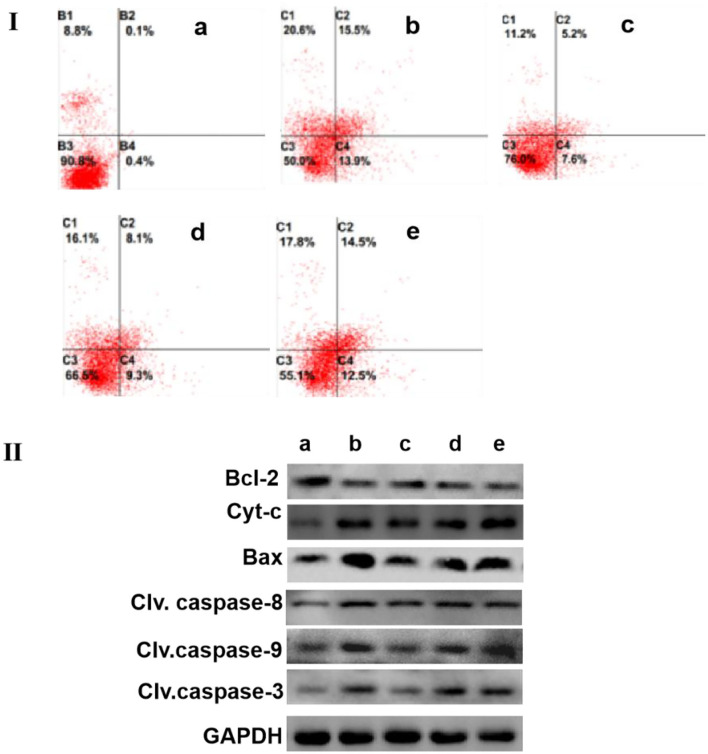
Figure 4.
SCIP improves the apoptosis of MCF-7. (I) The apoptosis level of MCF-7 cells was determined by flow cytometry analysis. MCF-7 Cells were cultured and incubated with 20 μg/ml of capecitabine and various concentrations of SCIP (27.8, 83.3, and 250 μg/ml) for 12 h, then, MCF-7 cells were stained with annexin V and PI, and flow cytometry was carried out to detect its apoptosis rate. (II) The expression of apoptosis-related proteins was investigated by western blots. MCF-7 cells were cultured and incubated with 20 μg/ml of capecitabine and various concentrations of SCIP (27.8, 83.3, and 250 μg/ml) for 12 h; After the treatment, the cells were collected to extract the total proteins and performed the western blot analysis to measure the expression of apoptosis proteins. a: Control group, MCF-7 cells were stimulated by PBS; b: Capecitabine group, MCF-7 cells were incubated by 20 μg/ml of capecitabine for 12 h; c: Low dose SCIP group, MCF-7 cells were incubated by 27.8 μg/ml of SCIP for 12 h; d: Moderate dose SCIP group, MCF-7 cells were incubated by 83.3 μg/ml of SCIP for 12 h; f: High dose SCIP group, MCF-7 cells were incubated by 250 μg/ml of SCIP for 12 h.

