Fig. 6.
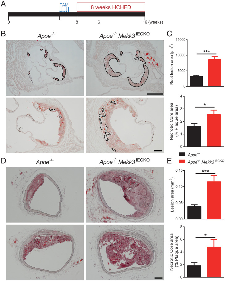
Endothelial MEKK3 knockout increases atherosclerotic plaque growth. (A) Experiment timeline: at 6 wk, Apoe−/− mice and Apoe−/− Mekk3iECKO mice were intraperitoneally injected tamoxifen for 5 consecutive d. From 8 wk, mice were fed HCHFD for 8 wk, then were euthanized for atherosclerosis analysis. (B) Representative Oil-Red-O (ORO) staining of aortic root. Outlines in the upper images indicate plaque lesion area and in the lower images indicate necrotic core area. (Scale bars, 500 μm [Upper] and 100 μm [Lower].) (C) Quantification of aortic root lesion area and necrotic core area for Apoe−/− mice (n = 10 male mice) and Apoe−/− Mekk3iECKO mice (n = 7 male mice). (D) Representative ORO staining of brachiocephalic artery. (Scale bar, 100 μm.) (E) Quantification of brachiocephalic artery lesion area and necrotic core area for Apoe−/− mice (n = 10 male mice) and Apoe−/− Mekk3iECKO mice (n = 7 male mice). Data represent mean ± SEM. *P < 0.05 and ***P < 0.001, calculated by unpaired t test.

