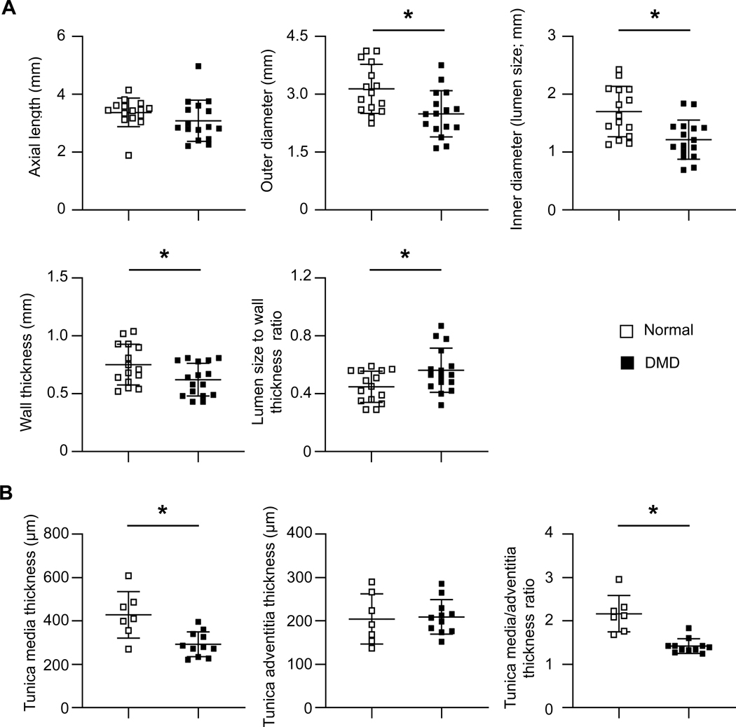
Figure 3. Quantitative characterization of normal and DMD dog femoral arteries by anatomic and histological measurements.
(A) Anatomic quantification of the axial length, outer diameter, inner diameter, wall thickness, and lumen size to wall thickness ration of normal (n=15) and DMD (n=16) dog femoral artery rings. (B) Histological quantification of the thickness of the tunica media and tunica adventitia, and the ratio of the tunica media thickness to tunica adventitia thickness of normal (n=7) and DMD (n=11) dog femoral arteries. Asterisk, significantly different between normal and DMD.

