Fig. 1.
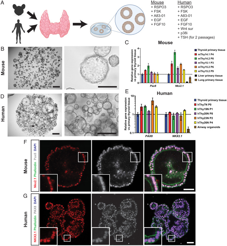
Establishment of thyroid follicular cell organoids from mouse and human origin. (A) Schematic representation of organoid derivation of TFCOs including medium components required for the establishment of murine TFCOs. Human TFCOs require additional components for establishment. (B) Brightfield images of murine TFCO cultures show cystic organoids grown in BME. (Scale bars, 1 mm [Left], 100 µm [Right].) (C) RT-qPCR analysis reveals similar to increased levels of Pax8 and Nkx2.1 expression in mouse TFCOs compared to primary thyroid tissue in early (fewer than four passages) and late (more than six passages) TFCOs while lung and liver tissue shows lower expression levels. n = 3. Error bars = SD. (D) Brightfield images of human TFCO cultures show cystic organoids grown in BME. (Scale bars, 1 mm [Left], 250 µm [Right].) (E) RT-qPCR analysis reveals similar to increased levels of PAX8 and NKX2.1 expression in human TFCOs compared to primary thyroid tissue in early (fewer than four passages) and late (more than six passages) TFCOs while lung tissue shows lower expression levels. n = 3. Error bars = SD. (F and G) Coexpression of thyroid transcription factors Nkx2.1/NKX2.1 (red) and Pax8/PAX8 (gray) in all cells in mouse (F) and human (G) TFCOs shows a pure population of TFCs in the organoids. Membrane staining was performed using Phalloidin (green), and nucleus staining using DAPI shows a single cell layer surrounding the lumen. (Scale bar, 100 µm.)

