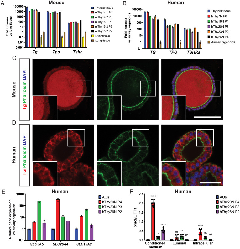
Fig. 4.
TFCOs express thyroid hormone machinery and secrete thyroid hormone basally. (A) RT-qPCR analysis reveals similar levels of Tg, Tpo, and Tshr expression in mouse TFCOs compared to primary thyroid tissue in early (fewer than four passages) and late (more than six passages) TFCOs, while liver tissue shows lower expression levels. Expression levels are relative to lung tissue expression levels. n = 3. Error bars = SD. (B) RT-qPCR analysis reveals slightly lower yet significant levels of TG and TPO and similar levels of TSHRa expression in human TFCOs compared to primary thyroid tissue in early (fewer than four passages) and late (more than six passages) TFCOs. Expression levels are relative to airway organoid expression levels. n = 3. Error bars = SD. (C) Mouse TFCs secrete Tg (red) toward the lumen in TFCOs similar to TFCs in follicles in vivo. Membrane staining was peformed using Phalloidin (green) and nucleus staining using DAPI (blue). (Scale bar, 100 µm.) (D) Human TFCs express TG (red) cytoplasmically in TFCOs. Membrane staining was performed using Phalloidin (green) and nucleus staining using DAPI (blue). (Scale bar, 50 µm.) (E) RT-qPCR analysis reveals expression of iodine transporters SLC5A5 and SLC26A4 and thyroid hormone transporter SLC16A2 in human TFCOs. Expression levels are relative to airway organoid (AO) expression levels. n = 3. Error bars = SD. (F) Human TFCOs in varying passages secrete variable yet measurable levels of free T3 (FT3) basally (conditioned medium) while apical secretion (luminal) is minimal or nondetectable. Some levels of FT3 are identified intracellularly (intracellular) yet at lower levels compared to basally. Each dot represents a separate expanded well of organoids measured. n = 5. Error bars = SD. ns, not significant, *P < 0.05, **P < 0.01, ****P < 0.0001 using two-way ANOVA with Tukey’s multiple comparisons to AOs.

