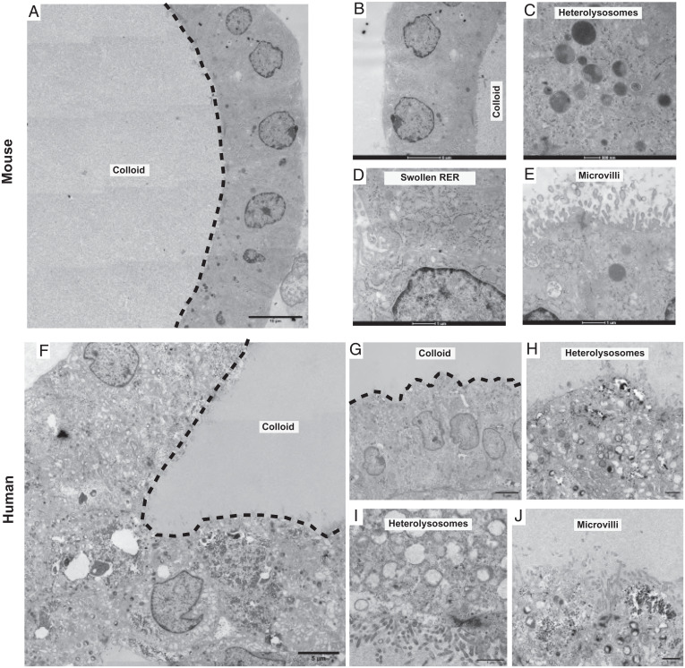
Fig. 5.
TFCs show short microvilli, swollen RER cisternae, and heterolysosomes and electrondense follicular lumens in TFCOs. (A) Overview image of transmission electron microscopy image reveals a variable in density lumen like the thyroid follicles show filled with potential colloid (dashed line indicates boundary of lumen) in mouse TFCOs. (Scale bar, 10 µm.) (B) Detailed zoom-in reveals a single cell layer of TFCs with cubic to columnar cells containing the nucleus in the basal area in mouse TFCOs. (Scale bar, 5 µm.) (C) Extensive heterolysosomes can be observed in mouse TFCOs indicating active transport of thyroglobulin to and from the lumen. (Scale bar, 500 nm.) (D) Swollen rough endoplasmic reticulum (RER) is present in mouse TFCOs indicative of TFCs. (Scale bar, 1 µm.) (E) Finger-like extensions of the plasma membrane can be observed as microvilli on the apical membrane of TFCs in mouse TFCOs. (Scale bar, 1 µm.) (F) Overview image of transmission electron microscopy reveals a variable in density lumen like the thyroid follicles show filled with potential colloid (dashed line indicates boundary of lumen) in human TFCOs. (Scale bar, 5 µm.) (G) Detailed zoom-in reveals a single cell layer with cubic to columnar cells containing the nucleus in the basal area of TFCs in human TFCOs. (Scale bar, 5 µm.) (H and I) Extensive heterolysosomes can be observed in human TFCOs, indicating active transport of thyroglobulin to and from the lumen. (Scale bars, 1 µm.) (J) Finger-like extensions of the plasma membrane can be observed as microvilli on the apical membrane of TFCs in human TFCOs. (Scale bar, 1 µm.)

