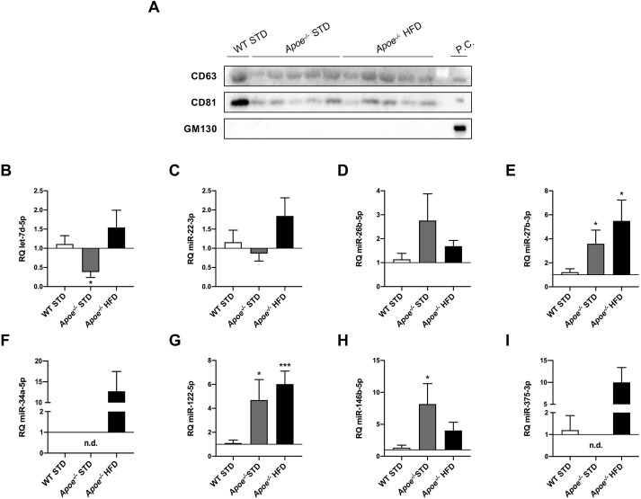
Fig. 7.
RT-qPCR analysis of miRNA expression in circulating extracellular vesicles. (A) Western blot analysis of markers to identify exosomes, such as CD63 and CD81, and GM130 as a cis-Golgi matrix protein used as a negative control, in WT STD (n=1), Apoe−/− STD (n=5) and Apoe−/− HFD (n=5) mice after 18 weeks on the diet. Huh7 cells were used as a positive control. (B-I) Comparison of let-7d-5p (B), miR-22-3p (C), miR-26b-5p (D), miR-27b-3p (E), miR-34a-5p (F), miR-122-5p (G), miR-146b-5p (H) and miR-375-3p (I) expression in WT STD (n=4-7), Apoe−/− STD (n=3-6) and Apoe−/− HFD (n=4-7) mice after 18 weeks on the diet. miR-16-5p was used as a control. Results are expressed as mean±s.e.m. Statistical significance was assessed by two-tailed unpaired Student's t-test, with the exception of miR-22-3p data, which were evaluated by unpaired non-parametric Mann–Whitney U test. *P<0.05 and ***P<0.001 versus WT STD mice. No statistics could be performed for miR-34a-5p and miR-375-3p data owing to undetermined values obtained in most WT STD and Apoe−/− STD liver samples. n.d., non-detected; P.C., positive control.

