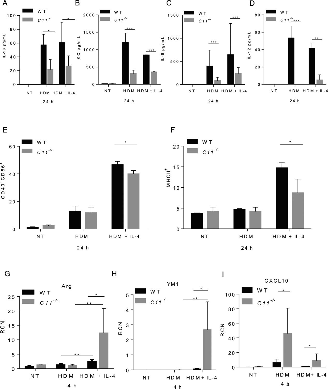
Fig. 2. Caspase-11−/− macrophages show reduced release of proinflammatory cytokines and reduced expression of costimulatory molecules post-HDM treatment.

Level of IL-1β released in the supernatants of WT or caspase-11−/− macrophages treated with 100 μg/mL of HDM after 24 h (A). Data represent the mean ± SD (n = 4) obtained from four independent experiments. Multiple t-tests performed for statistical analysis, * p < 0.05. Concentrations of keratinocyte-derived protein chemokine (KC) (B), IL-6 (C), and IL-12 (D) released in supernatants from WT and caspase-11−/− macrophages either un-treated (NT), treated with HDM (100 μg/mL), pre-stimulated with IL-4 (5 ng/mL) then treated with HDM and IL-4 for 24 h. Data represent the mean ± SD (n = 4) obtained from four independent experiments. Multiple t-tests performed for statistical analysis, ** p < 0.01, *** p < 0.001. Expression of costimulatory molecules CD40 and CD86 (E), and MHCII (F) by flow cytometry 24 h post- HDM treatment. Data represent the mean ± SD (n = 3) obtained from three independent experiments. Multiple t-tests performed for statistical analysis, * p < 0.05. WT and caspase-11−/− macrophages either NT, treated with HDM (100 μg/mL), pre-stimulated with IL-4 (5 ng/mL) then treated with HDM and IL-4 for 4 h. Expression of M2 (Arg1, YM1) or M1 (CXCL10) markers by RT-qPCR 4 h post-treatment (G, H & I). RCN on the Y axis represents relative copy number. Data represent the mean ± SD (n = 3) obtained from three independent experiments. Multiple t-tests performed for statistical analysis, * p < 0.05.
