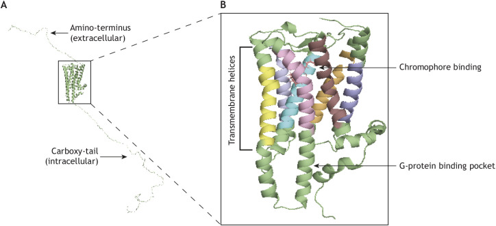
Fig. 1.
Structure of mammalian Opn4 based on squid rhodopsin. (A) 3D homology modeling of inactive mouse melanopsin based on a squid (Todarodes pacificus) rhodopsin template. Modeling was done in PyMOL (The PyMOL Moelcular Graphics System, v.1.2r3pre, Schrödinger, LLC). Mouse melanopsin boasts an exceptionally long carboxy-tail that heavily influences both activation and deactivation of melanopsin signaling. (B) An enhanced view of the transmembrane regions of mouse melanopsin, including chromophore binding. Each distinct transmembrane helix is represented in a different color. Upon isomerization of the chromophore by light, transmembrane movement of the transmembrane helices permits the binding of G-protein to intracellular loops two and three.

