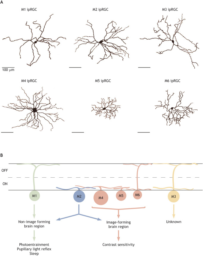
Fig. 2.
Overview of ipRGC subtype diversity. (A) Tracings of single M1–M6 ipRGCs. The properties of these cell types are summarized in Table 1. (B) A simplified schematic of M1–M6 stratification in the ON and OFF sublamina of the inner plexiform layer (IPL) depicting downstream targets. The projections of M3 have not been well-studied; however, they may innervate the superior colliculus (Zhao et al., 2014). The M1–M5 dye-filled cells were collected in the Schmidt laboratory, and the M6 morphology is reproduced from Quattrochi et al. (2019), with permission.

