Fig. 3.
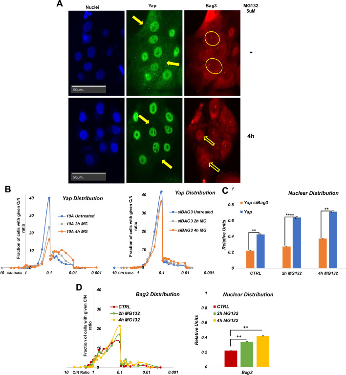
Effects of proteotoxic stress on nuclear localization of Bag3 and YAP. (A) Representative images of localization of YAP and Bag3 in the presence and absence of MG132. Filled arrows indicate the cytoplasmic content of YAP. Circles indicate nuclei with a low content of Bag3. Empty arrows indicate nuclei with a high content of Bag3. Images are representative of three experiments. (B) Distribution of cells in the population according to C/N ratio (cytoplasmic/nuclear localization) of YAP in control cells (left panel) and Bag3-depleted cells (right panel). Cells were treated on 96-well plates and fixed, and localization of YAP was assessed by immunofluorescence. Image acquisition and analysis were performed using the Hermes imaging system. Unlike control culture where MG132 caused redistribution of YAP in a large proportion of cells, in Bag3-depleted cells MG132 does not cause significant YAP redistribution. (C) Quantification of effects of MG132 treatment and Bag3 depletion on nuclear localization of YAP based on experiment in Fig. 3B. (D) Distribution of cells in the population according to C/N ratio of Bag3 in control cells (left panel). The treatments were performed as described in B. Right panel, quantification of effects of MG132 treatment on nuclear localization of Bag3 based on experiment in the left panel. **P≤0.01; ****P≤0.0001 (unpaired two-tailed t-test).

