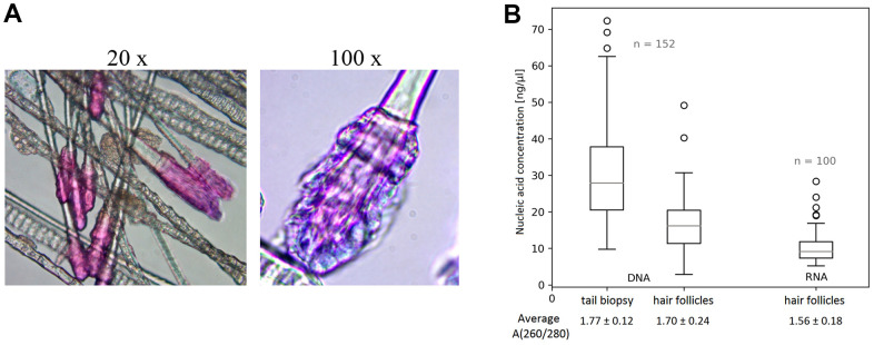
Figure 2.
Murine hair follicles as starting material. (A) Representative images of hairs highlighted with Giemsa – Romanowsky staining (and indicated magnification) showing intact hair follicles obtained by sampling with our vacuum collector. (B) Comparison of DNA isolation results from hair follicles obtained with our device and common tail biopsies, and yields of RNA isolated from hair follicles. The quantity and quality of all nucleic acid samples, in 30 μl solutions, were measured using a ND 1000 Spectrophotometer. Data are presented as boxplots showing median, maximum and minimum values, their quartile distribution, interquartile range, and outlier values.

