Figure 4.

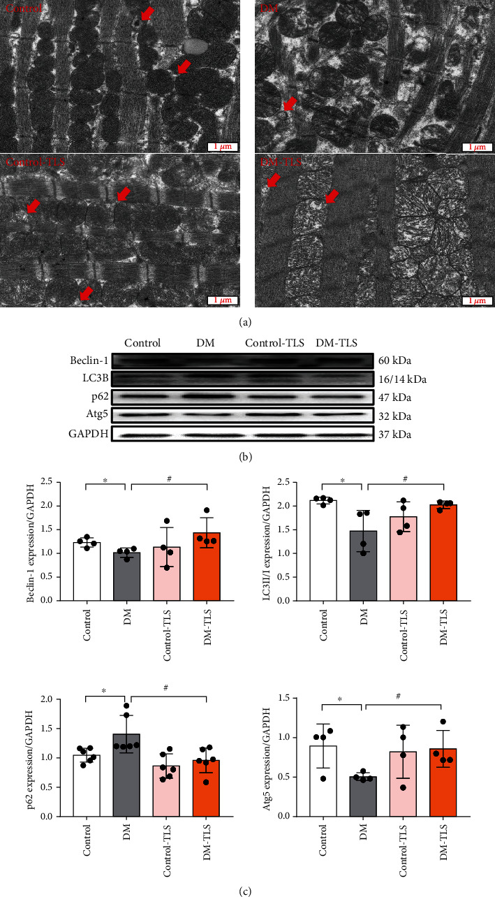
TLS activates autophagy in diabetic mice. (a) Representative transmission electron microscopy image. Red arrows mark autophagosomes. (b) Representative protein expression of Beclin-1, LC3B, p62, and Atg5. (c) Quantification of Beclin-1, LC3B, p62, and Atg5 protein expression. n = 4-6 per group. Means ± SD. ∗P < 0.05 compared to the control group, #P < 0.05 compared to the DM group.
