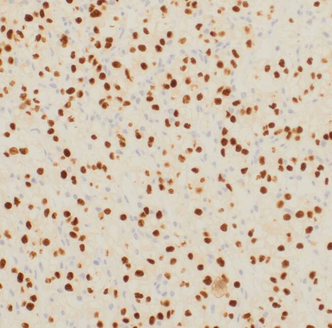
Fig. 3.

The immunohistochemistry for renal cell carcinoma from the sample taken from the tumor in the ampulla of Vater (case 1). It is renal cell cancer metastasis. The cells have clear cytoplasm. All the brown dots are the positive cancer cells/nuclei. Some nuclei are not taking the brown stain and they are vessels and fibroblasts. Positive staining was found for AE1/3,Pax 8, CD10, vimentin and EMA and these are consistent with renal cell cancer. The stains were negative for CK7, CK20, and TTF-1. EMA, epithelial membrane antigen.
