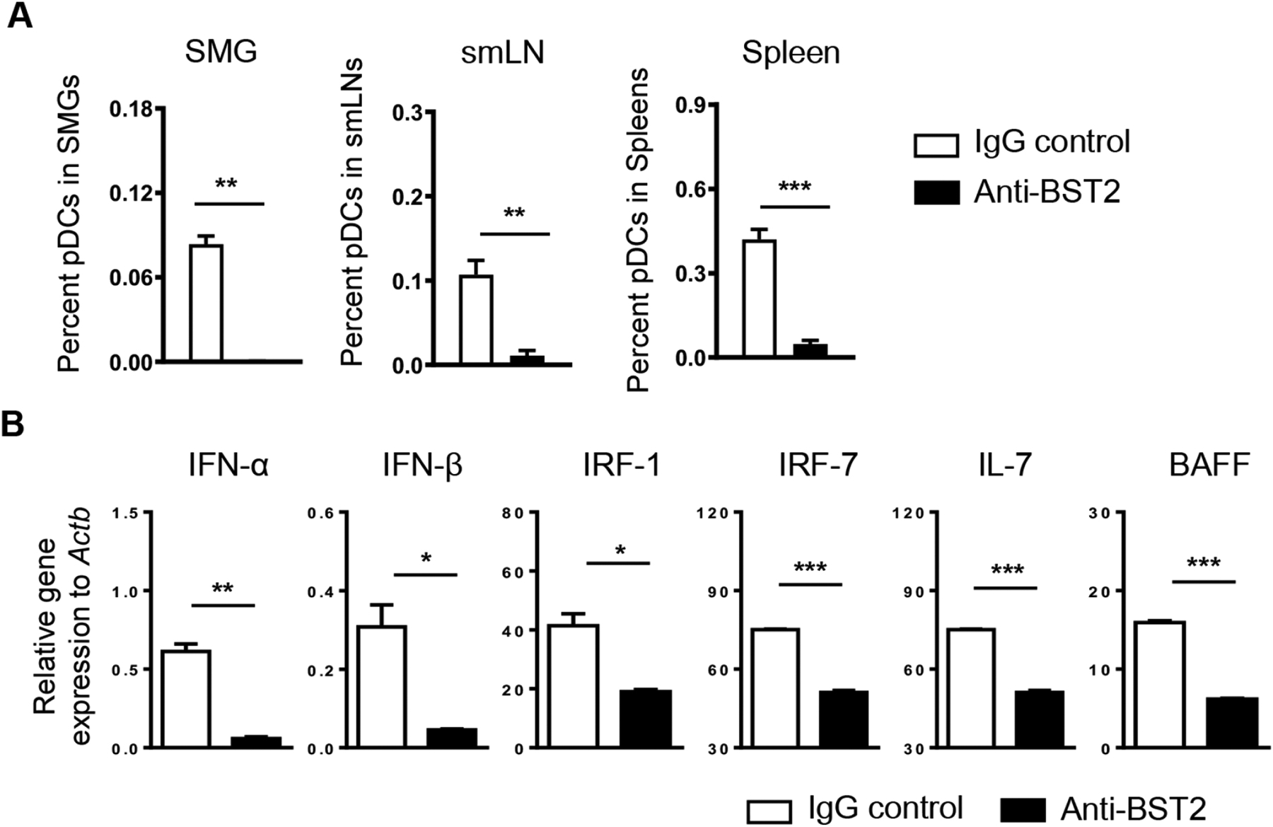
Figure 3. Administration of anti-BST2 antibody efficiently depletes pDCs and reduces expression of type I IFNs, type I IFN-responsive genes and SS-promoting cytokines in SMGs.

Anti-BST2 or isotype rat IgG was i.p. administered into 9-week-old female NOD mice on day 0 and day 2. The analyses were performed 24 hours after the last injection. (A) Percentages of pDCs (defined as CD11b−CD11cmidB220+Siglec-H+BST2+) among total SMG cells, SMG-draining lymph node, and spleen cells based on flow cytometric analysis. (B) Real-time PCR analysis of the mRNA levels of type I IFNs, type I IFN-responsive genes and SS-promoting cytokines in the SMGs. The results are presented relative to that of β-actin. All data are representative or the average of analyses of 3 mice for each group.
