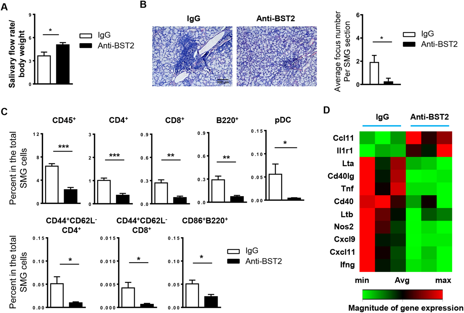
Figure 4. Administration of anti-BST2 antibody over a 3-week period reduces leukocyte infiltration of SMGs and improves salivary secretion in female NOD mice.

Anti-BST2 or isotype rat IgG was i.p. administered into 4-week-old female NOD mice, 3 times weekly for 3 weeks. All the analyses were performed when mice were 10 weeks of age. (A) Stimulated saliva flow rate normalized to body weight. (B) Images of H&E staining of SMG sections (scale Bar=100 μm). Bar graph shows the average number of leukocyte foci per SMG section. (C) Flow cytometric analysis of lymphocyte populations in the SMGs. (D) The clustergram of RT2 Profiler PCR Array results displaying gene expression levels with statistically significant changes upon anti-BST2 treatment. All data are representative or the average of analyses of 6 mice for each group.
