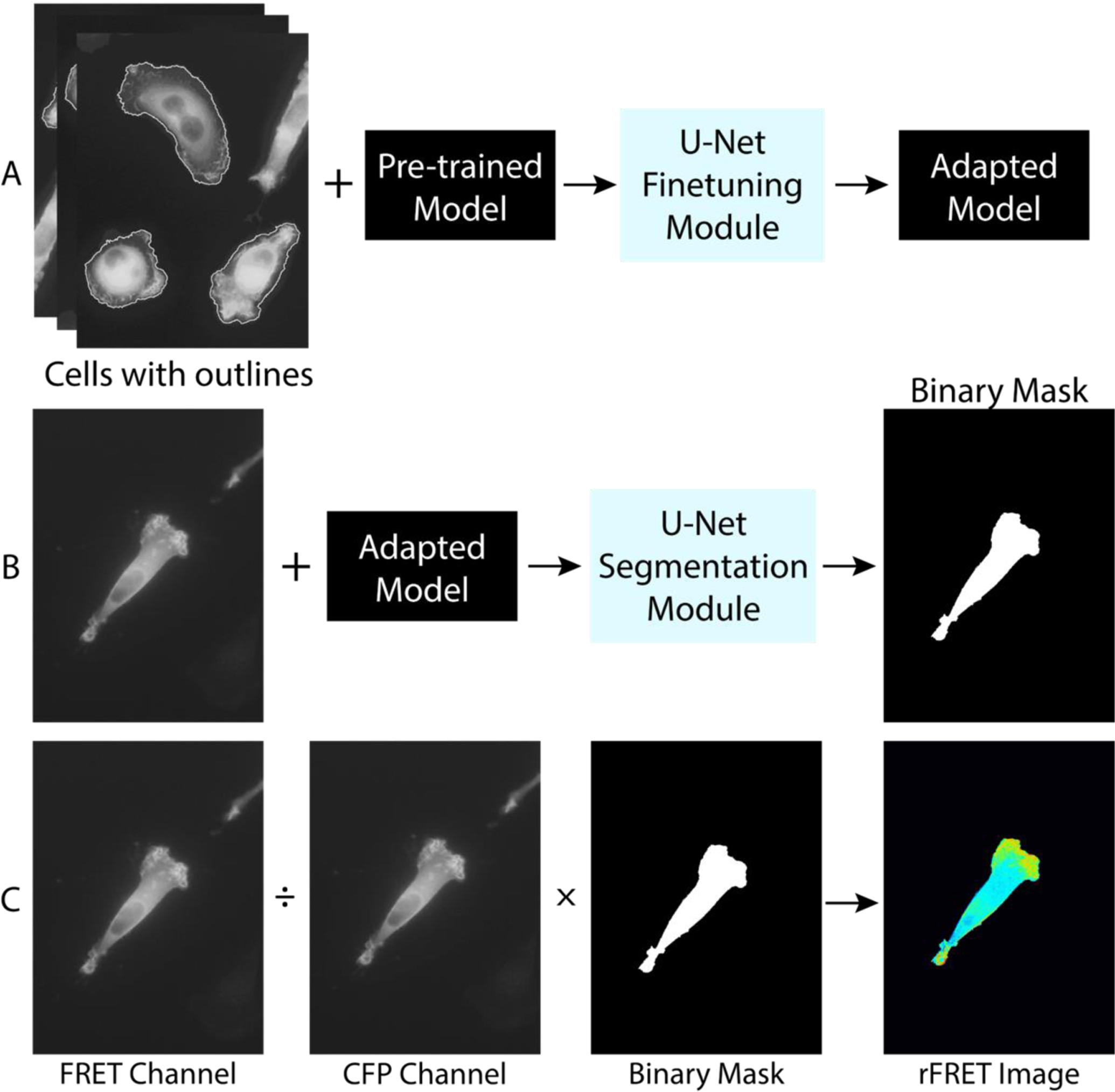
Figure 1. Illustration of a machine learning-assisted FRET technique.

A machine learning approach is adopted in cell segmentation prior to the ratiometric FRET calculation. A. Model training using a convolutional neural network (U-Net) package. Original images of cells with manually drawn outlines together with pre-trained model are input to the U-Net finetuning module. The output is the trained adapted model. B. Cell binary mask generation using the adapted model. The cell image from FRET channel and the adapted model are fed into the U-Net segmentation module. The output is a binary mask of the cell. Here, cell images from the FRET channel are used. C. Ratiometric FRET calculation. The ratiometric FRET image is obtained by dividing the image of the FRET channel from that of the CFP channel, and then segmented by the binary mask. The final image is then corrected for photobleaching.
