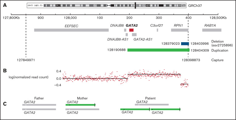
Figure 1.
Region of chromosome 3 showing copy number variation in the patient. (A) The captured region (3:127849971-128398873) is indicated by dashed lines, the duplication detected (3:128190688-128404309) is shown in green, and the deletion (3:128379023-128303996) is shown in blue. (B) The normalized read count mapping to the captured region. (C) The predicted arrangement of alleles in the parents and patient. The asterisk (*) indicates the position of GATA2, and the broken line denotes the site of the deletion.

