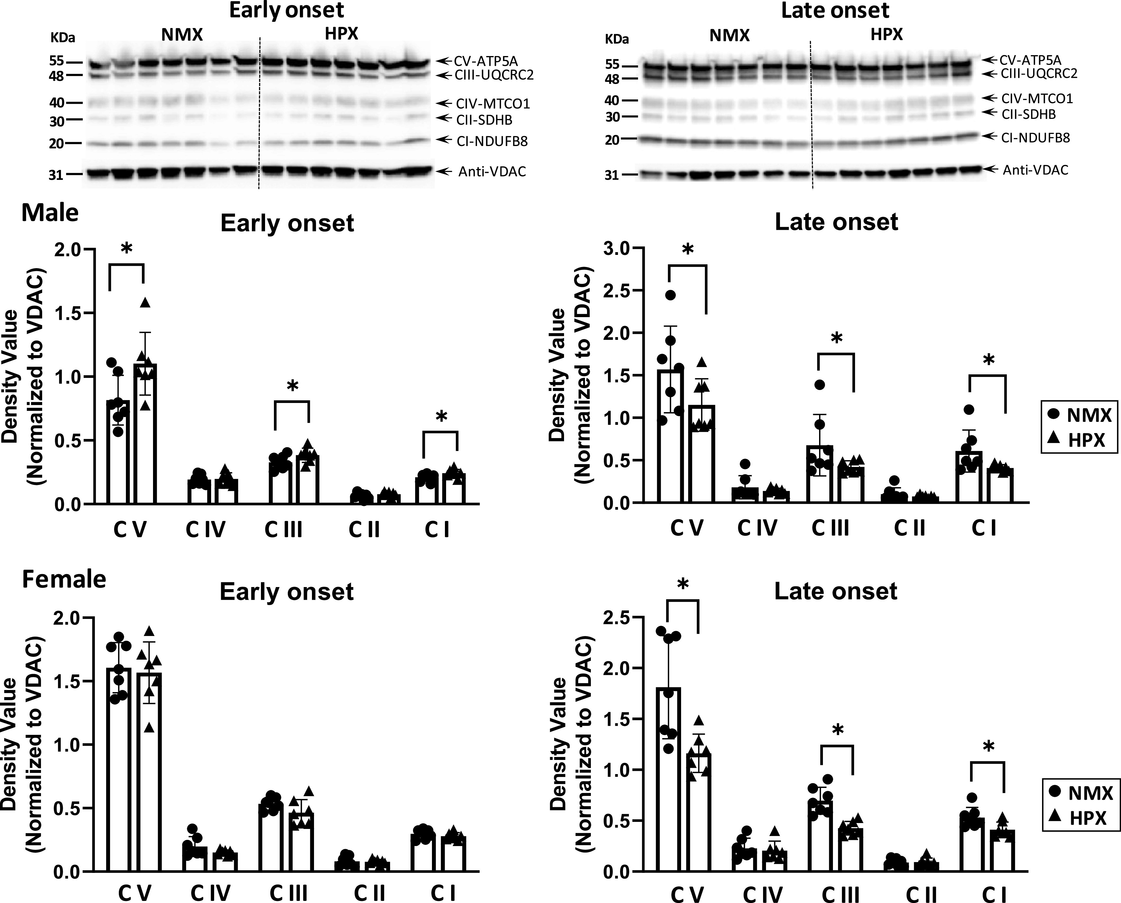
Figure 1.

Western immunoblot (top, male fetal hearts) and analysis (bottom) of mitochondrial complex (CI–V) subunit expression of isolated cardiac mitochondria of left ventricles. Male and female fetuses were exposed to normoxia (NMX, circles) and early-onset [25 days (25d) gestation, 39d duration] and late-onset (50d gestation, 14d duration) hypoxia (HPX, triangles). Protein expression was measured in fetal heart ventricles obtained at term (64d gestation). Density values are target band values normalized to VDAC loading controls. *Significant differences at P < 0.05 vs. NMX; n = 7 animals in each group. Representative blots identified Complex I–V subunits are NFUFB8, SDHB, UQCRC2, MITCO1, and ATP5a, respectively. VDAC, voltage-dependent anion channel.
