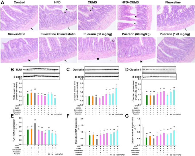
FIGURE 4.
Effect of puerarin on HFD/CUMS-induced small intestine mucosa. (A) Pathological damage of the small intestine was detected by H&E staining. indicates the thickness from the serosa to muscularis;
 shows that the villi were swollen and broken, and the arrangement of the lamina propria cells was completely disordered;
 indicates that the serosal layer was loosely arranged or fell off from the muscle layer. The magnification was 100 ×. The protein expression of TLR4 (B), occludin (C), and claudin-1 (D) was analyzed by Western blotting. β-actin was used as the internal reference. The densitometry was quantified using ImageJ software. The mRNA levels of TLR4 (E), occludin (F), and claudin-1 (G) were detected by real-time RT-PCR. β-actin was used as the housekeeping gene. Values are expressed as mean ± S.E.M (n = 3 for Western blotting and n = 4 for real-time RT-PCR). #
p < 0.05 and ##
p < 0.01 vs. the normal control group; *
p < 0.05 and **
p < 0.01 vs. the HFD/CUMS group.

