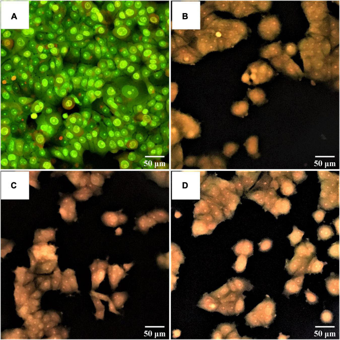
FIGURE 9.
Fluorescent photomicrographs of AO/PI-stained cells on MCF-7 cell lines treated with CFS of S. salivarius strains for 24 h. (A) Untreated cells, (B) MCF-7 cells treated with S. salivarius BP8, (C) MCF-7 cells treated with S. salivarius BP156, and (D) MCF-7 cells treated with S. salivarius BP160.

