FIGURE 5.
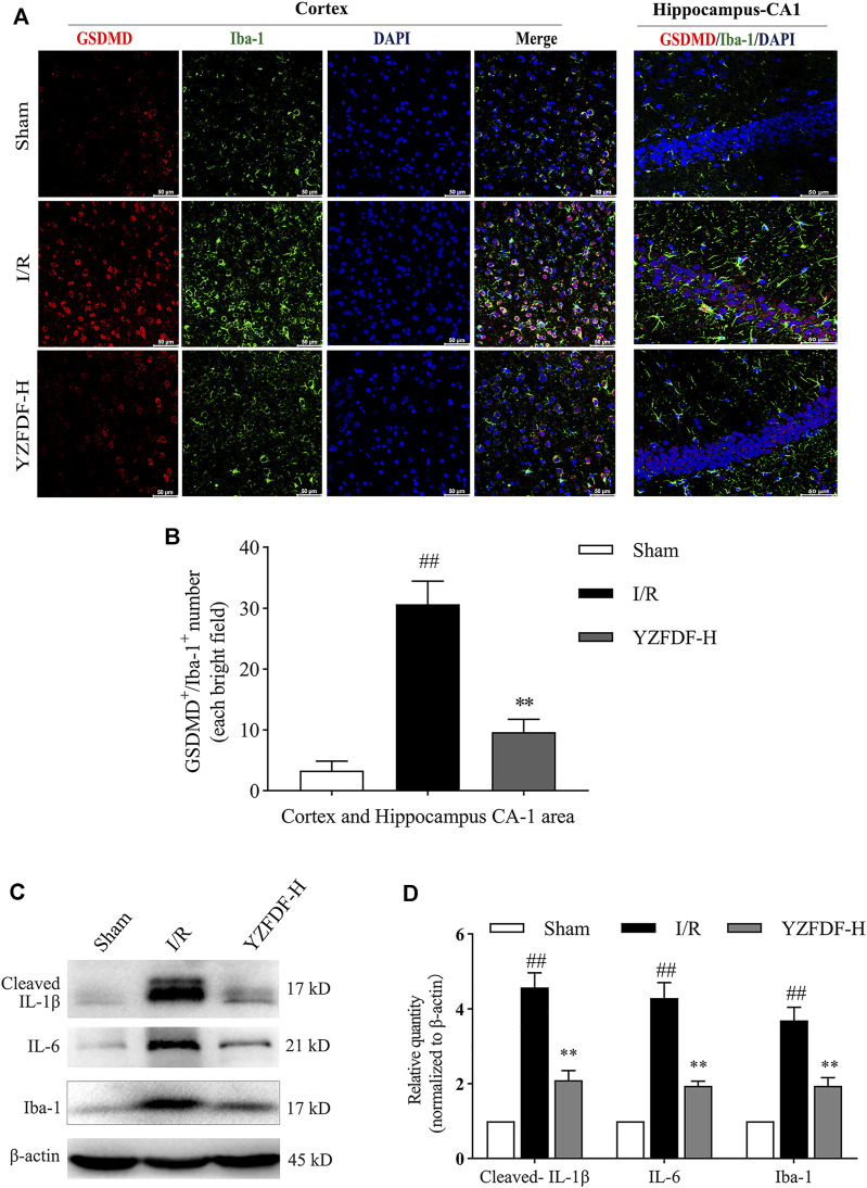
YZFDF blocked overactivation and pyroptosis of microglia and alleviated inflammatory responses at 24 h after cerebral I/R in rats. (A) Representative pictures of double immunofluorescence staining of GSDMD (red) colocalized with Iba-1 (green) in cortex and hippocampus-CA1 areas, scale bar = 50 μm. (B) Quantitative analysis of double-positive staining of GSDMD+/Iba-1+ cell number, n = 3. (C) Representative Western blots for cleaved-IL-1β, IL-6, and Iba-1. (D) Quantitative analysis of cleaved-IL-1β, IL-6, and Iba-1, n = 6. ## p < 0.01 vs. Sham group; ** p < 0.01 vs. I/R group.

