Figure 6.
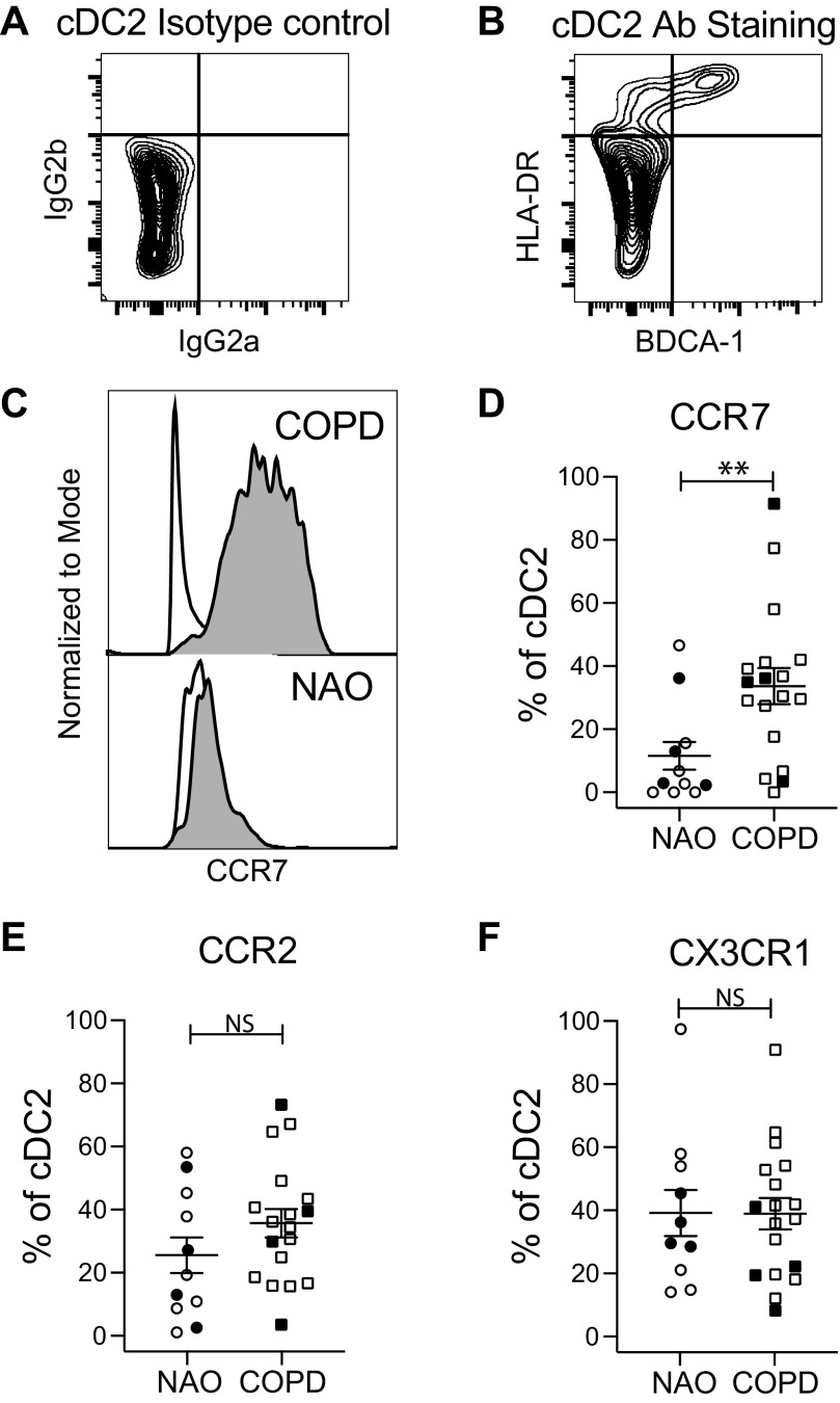
Identification of lung cDC2 and chemokine receptor expression. Single-cell suspensions of dispersed human lung tissue were stained with fixable live-dead stain and antibodies toward CD45, CD3, CD19, BDCA-1, HLA-DR, CCR7, CCR2, and CX3CR1. After gating on cells that were viable, CD45+, CD3−, and CD19−, cDC2 cells were identified by comparing an isotype control (A) with antibody staining (B) for HLA-DR and BDCA-1. C: representative staining showing the expression of CCR7 on cDC2 cells from subjects with NAO and those with COPD; white histogram, isotype control; gray histogram, CCR7 staining. D–F: compiled data from all subjects for the percentage of cDC2 expressing CCR7 (D), CCR2 (E), and CX3CR1 (F). Symbols indicate individual participants; circles, participants with NAO (n = 11) and squares, participants with COPD (n = 18). Lines represent the means ± SEM. The Mann–Whitney U test was used to determine significance; **P < 0.01. Closed symbols, current smokers; open symbols, former smokers. cDC2, conventional dendritic cell, type 2; COPD, chronic obstructive pulmonary disease; NAO, no airway obstruction; NS, not significant.

