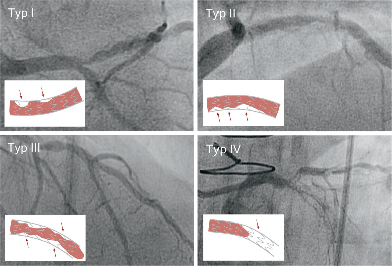
Figure 1:
Angiographic classification of in-stent restenosis (ISR) (adapted from [13])
Type I: Focal ISR with lesions ≤ 10 mm in length. Lesions can be located between neighboring stents, at the proximal or distal end of the stent, within the stent or at any combination of the above locations.
Type II: Diffuse ISR with lesions >10 mm in length. The lesions are limited to the stent, without extending beyond its ends.
Type III: Diffuse, proliferative ISR. The lesions are >10 mm in length and extend beyond the edges of the stent.
Type IV: ISR with total occlusion. Lesions with TIMI-0 flow*.
* TIMI-0 flow, no coronary perfusion; TIMI-1 flow, significantly slowed coronary blood flow; the categories range from TIMI-0 to TIMI-3.

