Figure 2:
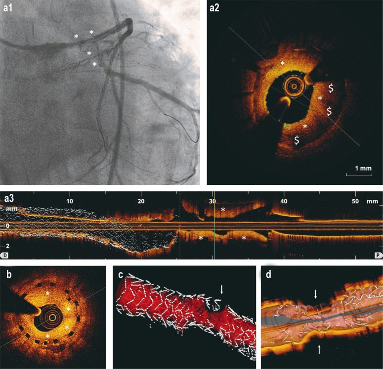
In-stent restenosis after DES implantation and implantation of bioresorbable coronary stents (scaffolds): selected coronary angiography and optical coherence tomography (OCT) findings
a) The images show an in-stent restenosis (*) (a1: coronary angiography; a2: cross-section in OCT; a3: longitudinal section in OCT) in the area of the left anterior descending artery (LAD) at three months after implantation of a DES in the main stem of the left coronary artery. The area of the lumen is 3.11mm2 (normal >8 mm2). The dark areas ($) are peri-strut low intensity areas (PSLIA) and suggestive of immature, rapidly growing neointima.
b) OCT in a 67-year-old man who presented without symptoms at the 6-month follow-up after implantation of a bioresorbable scaffold. The image shows a high-grade in-stent restenosis (*) in the circumflex artery (Cx).
c–d) 3D reconstructions of in-stent restenoses (arrows) in OCT. In Figure c, the blood vessel lumen is marked in red.
DES, drug-eluting stent

