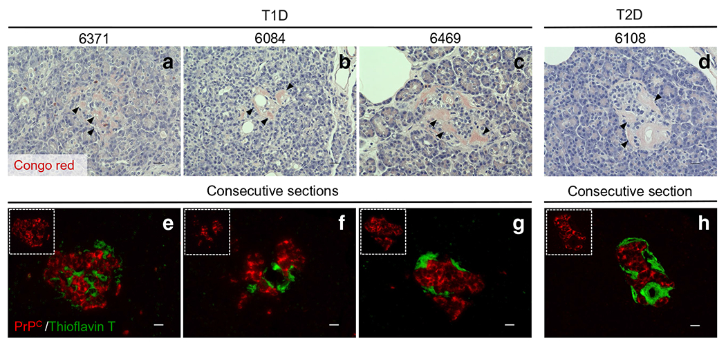
Fig. 5.

Assessment of possible interactions between PrPC and protein aggregates in type 1 and type 2 diabetic donors. Among our 12 type 1 diabetic organ donors, three cases displayed amyloid-like plaques. In these three cases, we evaluated whether PrPC co-localised with these islet-associated protein aggregates. (a)–d) The pathology stain, Congo Red, was used to identify amyloid/protein aggregates in the three type 1 diabetic donors as well as one type 2 diabetic donor (positive control). Black arrows show positivity for amyloid-like plaques (pink colour) in pancreatic islets. (e)–h) To visualise amyloid-like plaques and PrPC expression together, consecutive slides were stained with thioflavin T (green) and PrPC (red). We found no colocalisation of PrPC-positive islet cells with the thioflavin T regions in either the type 1 or the type 2 diabetic pancreas samples. Scale bars, 20 μm. T1D, type 1 diabetic; T2D, type 2 diabetic
