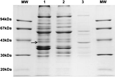
FIG. 1.
Analysis of B. clarridgeiae flagellin preparation by SDS-PAGE and Coomassie brilliant blue staining. Lanes: MW, molecular mass marker; 1, whole-cell lysates of B. clarridgeiae; 2, cellular debris after shearing off the flagella; 3, crude flagellar protein. The arrow indicates the position of the 41-kDa band identified as the FlaA protein.

