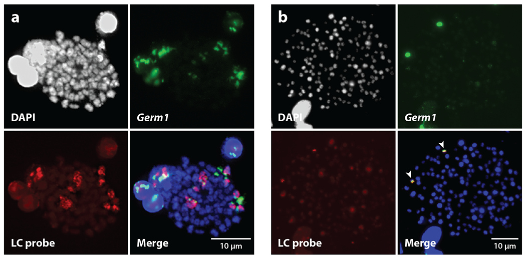
Figure 3.

Differences between lamprey germline and somatic karyotypes. (a) A meiotic metaphase I chromosome spread. (b) A mitotic metaphase spread generated from a 16-day-old embryo. Chromosomes were stained with DAPI and labeled with fluorescence in situ hybridization probes to the Germ1 repeat (green) and a laser capture (LC; red) probe, generated from eliminated chromatin (41, 120). Both Germ1 and LC probes hybridize with multiple tetrads in meiotic metaphase I, whereas signals are observed on only one pair of mitotic chromosomes (indicated with arrows). Hybridization of the LC probe to somatic cells yields weaker signals that are only slightly brighter than background fluorescence, in contrast to hybridization patterns on meiotic chromosomes.
