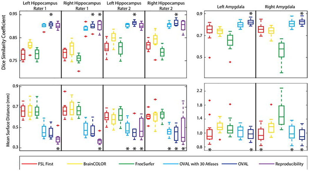
Figure 1:

Quantitative segmentation results for the whole hippocampus and amygdala. OVAL outperforms all other segmentation techniques in terms of DSC for the left hippocampus in both raters, the right hippocampus in rater 2, and the left and right amygdala (p<0.05, *). OVAL outperforms all other techniques for the right hippocampus of rater 1 except human reproducibility, which performs comparably statistically. Human reproducibility outperforms all other techniques in MSD for the left and right hippocampus for rater 1 (p<0.05, *). OVAL and OVAL-30 outperform all other automated techniques for those structures. OVAL, OVAL-30, and human reproducibility perform statistically comparable for the left and right hippocampus for rater 2 and outperform all other techniques (p<0.05, *). OVAL and FSL FIRST perform statistically similarly for the left amygdala and outperform all other segmentation approaches (p<0.05, *). OVAL, OVAL-30, and FSL FIRST perform statistically similarly for the right amygdala and outperform all other segmentation approaches (p<0.05, *).
