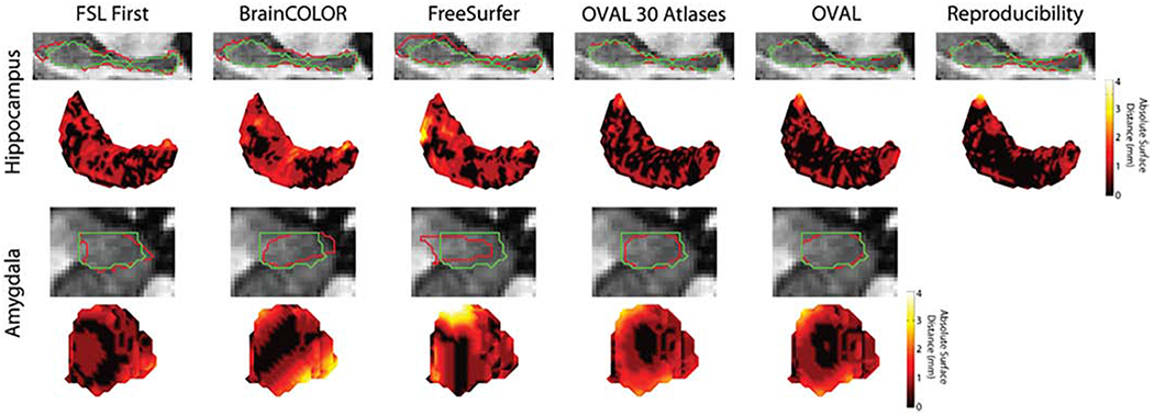
Figure 5:

Median qualitative segmentation results for the whole hippocampus and amygdala; red represents the estimated segmentation and green is the manual label. FSL FIRST, BrainCOLOR, and FreeSurfer all showed large surface distances up to 4mm for both the hippocampus and amygdala. OVAL and OVAL-30 were typically within 1mm distance on the hippocampus, though OVAL produced more consistent results than OVAL with 30 atlases. On the amygdala, OVAL and OVAL-30 captured the overall contour of the amygdala.
